
| 別品名 |
rabbit anti-ZO-1 antibody, ZO 1, ZO1, Tight junction protein ZO-1, Tight junction protein 1, Zona occludens protein 1, Zonula occludens protein 1, TJP1
|
| 抗原部位 |
Internal
|
| 種由来 |
Human
|
| 標識物 |
Unlabeled
|
| 精製度 |
Affinity Purified
|
| 適用 |
Western Blot
Enzyme Linked Immunosorbent Assay
Immunohistochemistry
Immuno Fluorescence
Flow Cytometry
|
| 免疫動物 |
Rabbit
|
| 交差種 |
Human
Mouse
|
| GENE ID |
7082
|
| Accession No.(Gene/Protein) |
NP_003248, Q07157
|
| Gene Symbol |
TJP1
|
| 形状 |
液状
|
| その他 |
[Buffer]0.02 M Potassium Phosphate, 0.15 M Sodium Chloride, pH 7.2
|
| 参考文献 |
[Pub Med ID]32715761
|
| [注意事項] |
濃度はロットによって異なる可能性があります。メーカーDS及びCoAからご確認ください。
|
|
※サムネイル画像をクリックすると拡大画像が表示されます。
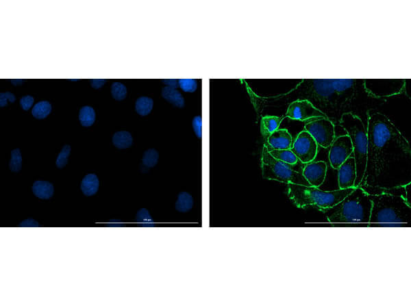
Immunofluorescence microscopy of Anti ZO 1 in Caco 2 cells using FITC conjugated Fluorescent TrueBlotR anti rabbit IgG (p/n 18 0216 32) for detection. Caco 2 cells were fixed with 4% PFA, blocked (5% mouse serum/0.3% Triton X 100 in 1X PBS) for 1hr, then incubated with 15ug/mL of anti ZO 1 primary antibody (Cat. No. 600 401 GU7) at 4C overnight. Following 3 washes in 1X PBS for 5min each, 5ug/mL of FITC conjugated Fluorescent TrueBlotR anti rabbit IgG was added and allowed to incubate for 1hr at room temperature. Nuclei were counterstained with DAPI present in mounting medium. Predicted cell localization is cell membrane and cell junctions. Image taken at 40X magnification. (Right) Merged DAPI (blue)/ZO 1 (green), image shown (Left) secondary antibody only.
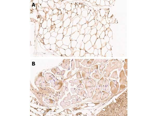
Immunohistochemistry of Rabbit anti-ZO-1 antibody. Tissue: mouse adipose tissue. Fixation: formalin fixed paraffin embedded. Epitope retrieval: heat induced (HIER). Primary antibody: ZO-1 antibody at 1:100 [A] and 1:200 [B] for 1 h at RT. Localization: ZO-1 will stain cell-cell junctions. Visualized with WARP RED on MACH 4 universal AP polymer detection system.
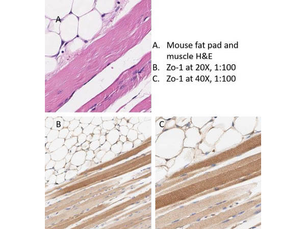
Immunohistochemistry of Rabbit Anti-ZO-1 antibody. Tissue: mouse adipose tissue and muscle. Fixation: formalin fixed paraffin embedded. Antigen retrieval: heat induced (HIER) using Citrate Buffer for 20min. Primary antibody: ZO-1 antibody at 1:100 for 30min at RT. Secondary Antibody: Anti-Rabbit Poly-HRP-IgG Ready-to-Use for 8min at RT. Localization: ZO-1 will stain cell-cell junctions. Staining: DAB. Counter Stain: Hematoxylin.
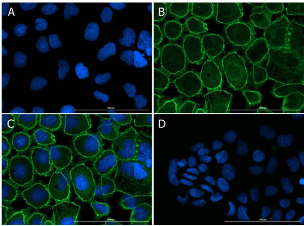
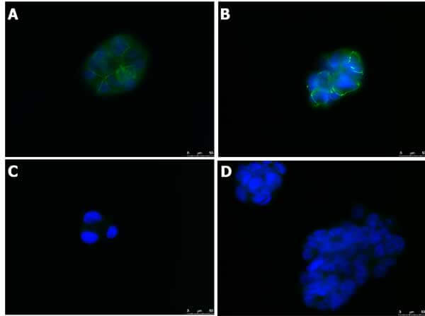
Immunofluorescence Microscopy of Rabbit anti-ZO-1 antibody. Tissue: CaCO2. Fixation: 4% PFA. Permeabilization: 0.3%Triton X-100. Primary antibody: ZO-1 antibody at 15μg/mL overnight at 2-8°C. Secondary antibody: Donkey Anti-Rabbit IgG DyLight? 488 Conjugated Preadsorbed (p/n 611-741-127) at 5μg/mL for 1 h at RT. Localization: membrane. Staining: (A)DAPI. (B)DyLight488. (C)Merge A-B. (D) Secondary Only.
Immunofluorescence Microscopy of Rabbit anti-ZO-1 antibody. Tissue: Caco2. Fixation: 0.5% PFA [A,C]. 0.5% MeOH [B,D]. Antigen retrieval: not required. Primary antibody: ZO-1 antibody at 10 ug/mL for 1 h at RT. Secondary antibody: Anti-RABBIT IgG DyLightTM 488 Conjugated Preadsorbed (p/n 611-741-127) at 5 ug/ml for 1 h at RT. Localization: (1) most epithelial cell junctions; (2) both in endothelial cells and the highly specialized epithelial junctions of renal and Sertoli cells. Staining: Target as green fluorescent signal with DAPI (blue) nuclear counterstain.
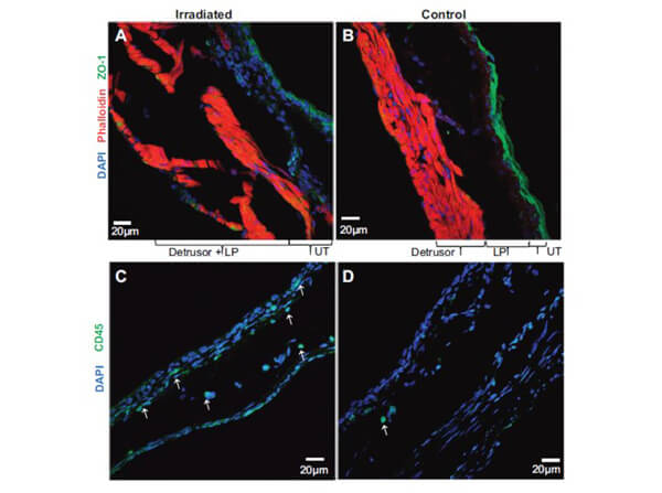
Immunofluorescence of ZO-1 and CD45. Representative confocal images of irradiated group (A) and controls (B) demonstrate a dramatic reduction of green fluorescence for ZO-1 in urothelium layer of irradiated group. Bladder sections were labeled with anti-ZO-1 antibodies and secondary donkey antibody Alexa 488 to localize the tight junction protein ZO-1 and with rhodamine-phalloidin to label the actin cytoskeleton (red) and DAPI to label the nuclei. Decreased expression of ZO-1 in urothelium layer of irradiated group was associated with an increase in CD45-positive, green fluorescent leukocytes (indicated by white arrows) in urothelium and lamina propria (C) compared with controls (D). Increased number of CD45-positive leukocytes indicates infiltration in irradiated bladder beyond the resident CD45 leukocytes spotted at one place in control section. Scale bar indicates magnification in different panels. Image Courtesy of Nishant Singh, Department of Urology, University of Pittsburgh, Pennsylvania.
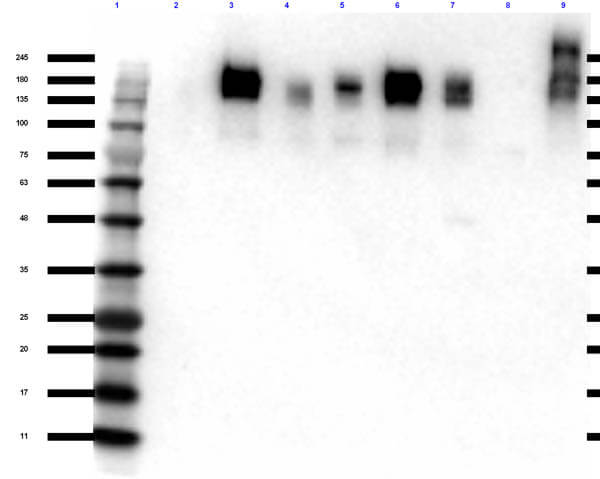
Western Blot of Rabbit Anti-ZO-1 Antibody. Lane 1: Opal Pre-stained MW ladder (p/n MB-210-0500). Lane 2: WM-115 Whole Cell Lysate (p/n W09-001-GY8). Lane 3: A549 Whole Cell Lysate (p/n W09-001-372). Lane 4: HeLa Whole Cell Lysate (p/n W09-000-364). Lane 5: HeLa Whole Cell Lysate CCCP Stimulated (p/n W09-001-GZ0). Lane 6: PC-3 Whole Cell Lysate (p/n W09-001-GV6). Lane 7: SK-OV-3 Whole Cell Lysate (p/n W09-001-GX9). Lane 8: Mouse Testis Lysate (p/n W10-000-GZ2). Lane 9: Rat Testis Lysate (p/n W12-000-GZ3). Load: 10 ug per lane. Primary antibody: ZO-1 antibody at 1:1000 for overnight at 4C. Secondary antibody: rabbit secondary HRP antibody (p/n 611-103-122) at 1:70,000 for 1 hr at RT. Block: 5% BLOTTO (p/n B501-0500) for 30 min at RT. Predicted/Observed size: ~187, 195 kDa for ZO-1.
|
|
|
|
Immunofluorescence microscopy of Anti ZO 1 in Caco 2 cells using FITC conjugated Fluorescent TrueBlotR anti rabbit IgG (p/n 18 0216 32) for detection. Caco 2 cells were fixed with 4% PFA, blocked (5% mouse serum/0.3% Triton X 100 in 1X PBS) for 1hr, then incubated with 15ug/mL of anti ZO 1 primary antibody (Cat. No. 600 401 GU7) at 4C overnight. Following 3 washes in 1X PBS for 5min each, 5ug/mL of FITC conjugated Fluorescent TrueBlotR anti rabbit IgG was added and allowed to incubate for 1hr at room temperature. Nuclei were counterstained with DAPI present in mounting medium. Predicted cell localization is cell membrane and cell junctions. Image taken at 40X magnification. (Right) Merged DAPI (blue)/ZO 1 (green), image shown (Left) secondary antibody only.
|
|
|
| メーカー |
品番 |
包装 |
|
RKL
|
600-401-GU7
|
100 UG
|
※表示価格について
| 当社在庫 |
なし
|
| 納期目安 |
約10日
|
| 保存温度 |
-20℃
|
|

