
| 別品名 |
rabbit anti-BCL3 antibody, rabbit anti-BCL-3 Antibody, BCL 3, B-cell lymphoma 3 protein, B-cell lymphoma 3 protein Antibody
|
| 抗原部位 |
C-Terminus
|
| 種由来 |
Human
|
| 標識物 |
Unlabeled
|
| 精製度 |
Affinity Purified
|
| 適用 |
Western Blot
Enzyme Linked Immunosorbent Assay
Immuno Fluorescence
|
| 免疫動物 |
Rabbit
|
| 交差種 |
Human
|
| GENE ID |
602
|
| Accession No.(Gene/Protein) |
NP_005169, P20749
|
| Gene Symbol |
BCL3
|
| 形状 |
液状
|
| その他 |
[Buffer]0.02 M Potassium Phosphate, 0.15 M Sodium Chloride, pH 7.2
|
| [注意事項] |
濃度はロットによって異なる可能性があります。メーカーDS及びCoAからご確認ください。
|
|
※サムネイル画像をクリックすると拡大画像が表示されます。
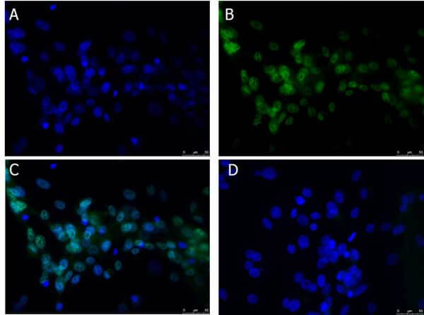
Immunofluorescence Microscopy of Rabbit anti BCL3 antibody. Tissue: U 87 MG cells. Fixation: 4% PFA. Antigen retrieval: not required. Primary antibody: BCL3 antibody at 1 ug/mL overnight at 4C. Secondary antibody: Donkey Anti Rabbit IgG DyLightTM 488 (p/n 611 741 127) at 5 ug/ml for 2 h at RT. Localization: BCL3 is subcellularly localized in the cytosol, nucleoplasm, midbody and vesicles. Staining: BCL3 as green fluorescent signal with DAPI (blue) nuclear counterstain.
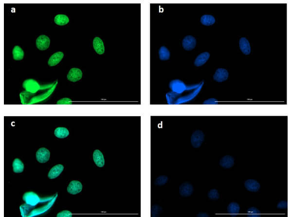
Immunofluorescence microscopy of BCL3 in Caco-2 cells using FITC-conjugated Fluorescent TrueBlotR anti-rabbit IgG for detection. Caco-2 cells were fixed with 4% PFA, blocked (5% mouse serum/.3% Triton X-1 in 1X PBS ) for 1 hr, then incubated with 15 ug/mL of anti-BCL3 primary antibody (Cat. No. 6-41-GU4) at 4C overnight. Following 3 washes in 1X PBS for 5 min each, 5 ug/mL of FITC-conjugated Fluorescent TrueBlotR anti-rabbit IgG was added and allowed to incubate for 1 hr at room temperature. Nuclei were counterstained with DAPI present in mounting medium. The predicted main localization is nucleoplasm. Additional localization in some cell types includes vesicles and midbody. (a) BCL3 (b) DAPI (c) merged DAPI/BCL3 (d) secondary antibody only. Image taken at 4X magnification.
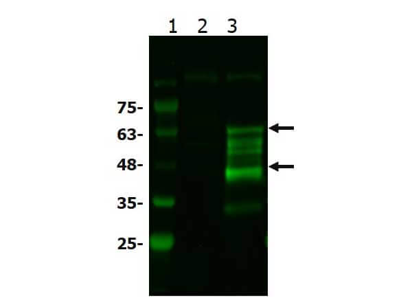
Western Blot of Rabbit anti-BCL3 antibody. Lane 1: MW ladder (p/n MB-21-5). Lane 2: HEK293T WCL. Lane 3: BCL3 overexpressing HEK293T lysate. Load: 1 ug per lane. Primary antibody: BCL3 antibody at 1:1 for overnight at 4C. Secondary antibody: goat anti-rabbit secondary Antibody DyLightTM 488 antibody (p/n 611-141-122) at 1:2, for one hour at RT. Block: BlockOut blocking buffer (p/n MB-73) one hour at RT. Predicted/Observed size: 48 kda. Other band(s): BCL3 processing caused by ubiquitination, phosphorylations.
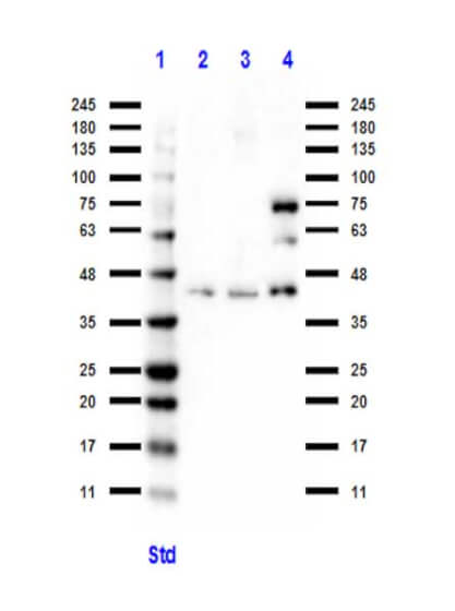
Western Blot of Rabbit anti-BCL3 antibody. Lane 1: MW ladder (p/n MB-21-5). Lane 2: U-87 MG WCL. Lane 3: C2C12 WCL (p/n W1-1-GL7). Lane 4: NIH/3T3 WCL (p/n W1--358). Load: 1 ug per lane. Primary antibody: BCL3 antibody at 1:1 for overnight at 4C. Secondary antibody: goat anti-rabbit secondary HRP antibody (p/n 611-13-122) at 1:7, for one hour at RT. Block: 1% Casein (p/n MB-81-1) (PVDF) blocking buffer one hour at RT. Predicted/Observed size: 47 kda. Other band(s): BCL3 processing caused by ubiquitination, phosphorylations.
|
|
|
|
Immunofluorescence Microscopy of Rabbit anti BCL3 antibody. Tissue: U 87 MG cells. Fixation: 4% PFA. Antigen retrieval: not required. Primary antibody: BCL3 antibody at 1 ug/mL overnight at 4C. Secondary antibody: Donkey Anti Rabbit IgG DyLightTM 488 (p/n 611 741 127) at 5 ug/ml for 2 h at RT. Localization: BCL3 is subcellularly localized in the cytosol, nucleoplasm, midbody and vesicles. Staining: BCL3 as green fluorescent signal with DAPI (blue) nuclear counterstain.
|
|
|
| メーカー |
品番 |
包装 |
|
RKL
|
600-401-GU4
|
100 UG
|
※表示価格について
| 当社在庫 |
なし
|
| 納期目安 |
約10日
|
| 保存温度 |
-20℃
|
|

