|
※サムネイル画像をクリックすると拡大画像が表示されます。
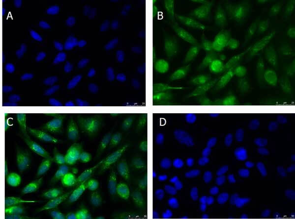
Immunofluorescence Microscopy of Rabbit anti-xCT antibody. Tissue: PC3 cells. Fixation: 100% MeOH. Antigen retrieval: not required. Primary antibody: xCT antibody at 10 μg/mL overnight at 4?C. Secondary antibody: Donkey Anti-Rabbit IgG DyLight? 488 (p/n 611-741-127) at 5 ug/ml for 2 h at RT. Localization: xCT is localized on the cell membrane and vesicles. Staining: xCT as green fluorescent signal with DAPI (blue) nuclear counterstain.
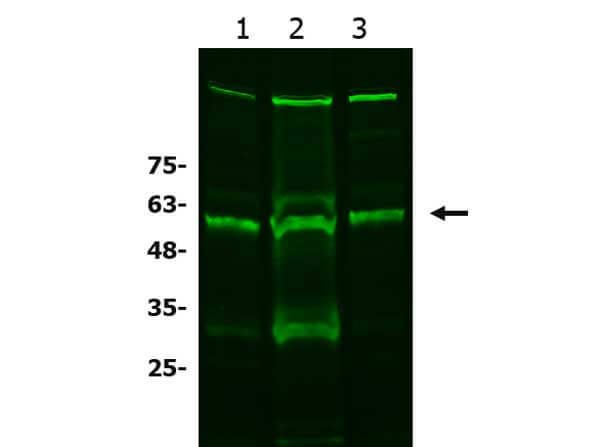
Western Blot of Rabbit anti-xCT antibody. Lane 1: A549 WCL (p/n W09-001-372). Lane 2: HCT-116 WCL (p/n W09-001-GM4). Lane 3: HeLa WCL (p/n W09-000-364). Load: 10 μg per lane. Primary antibody: xCT antibody at 1:1000 for overnight at 4°C. Secondary antibody: donkey anti-rabbit secondary DyLight?488 antibody (p/n 611-741-127) at 1:20,000 for one hour at RT. Block: BlockOut blocking buffer (p/n MB-073) one hour at RT. Predicted/Observed size: 56 kda. Other band(s): xCT processing caused by dimerization, glycosylation, and/or phosphorylation.
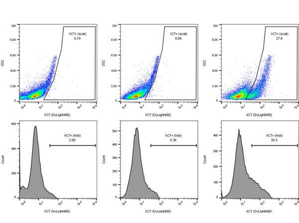
Flow Cytometry of rabbit anti-xCT antibody. Cells: breast carcinoma cells Primary antibody: xCT antibody at 1.0 μg/mL for one hour at 4°C. Secondary antibody: Donkey anti-Rabbit IgG Dylight?488 Antibody p/n (611-741-127) at 1 ug/ml in 200 ul for one hour on ice.
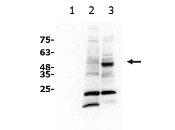
Western Blot of Rabbit anti-xCT antibody. Lane 1: recombinant BCL3 (unrelated negative control). Lane 2: NIH 3T3 WCL (p/n W10-000-358). Lane 3: A549 WCL (p/n W09-001-372). Load: 10 μg per lane. Primary antibody: xCT antibody at 1:1000 for overnight at 4°C. Secondary antibody: goat anti-rabbit secondary HRP antibody (p/n 611-103-122) at 1:20,000 for one hour at RT. Block: 5% BSA blocking buffer one hour at RT. Predicted/Observed size: 56 kda. Other band(s): xCT processing caused by dimerization, glycosylation, and/or phosphorylation.
|
|
|
|
Immunofluorescence Microscopy of Rabbit anti-xCT antibody. Tissue: PC3 cells. Fixation: 100% MeOH. Antigen retrieval: not required. Primary antibody: xCT antibody at 10 μg/mL overnight at 4?C. Secondary antibody: Donkey Anti-Rabbit IgG DyLight? 488 (p/n 611-741-127) at 5 ug/ml for 2 h at RT. Localization: xCT is localized on the cell membrane and vesicles. Staining: xCT as green fluorescent signal with DAPI (blue) nuclear counterstain.
|
|
| 別品名 |
rabbit anti-xCT antibody, SLC7A11, Cystine/glutamate transporter, Amino acid transport system xc-, Calcium channel blocker resistance protein CCBR1, Solute carrier family 7 member 11
|
| 交差種 |
Human
|
| 適用 |
Western Blot
Enzyme Linked Immunosorbent Assay
Immuno Fluorescence
Flow Cytometry
|
| 免疫動物 |
Rabbit
|
| 抗原部位 |
Internal
|
| 標識物 |
Unlabeled
|
| 精製度 |
Affinity Purified
|
| GENE ID |
23657
|
| Accession No.(Gene/Protein) |
NP_055146.1, Q9UPY5
|
| Gene Symbol |
SLC7A11
|
| その他 |
[Buffer]0.02 M Potassium Phosphate, 0.15 M Sodium Chloride, pH 7.2
|
| 参考文献 |
[Pub Med ID]37444594
|
| [注意事項] |
濃度はロットによって異なる可能性があります。メーカーDS及びCoAからご確認ください。
|
|
| メーカー |
品番 |
包装 |
|
RKL
|
600-401-GU3
|
100 UG
|
※表示価格について
| 当社在庫 |
なし
|
| 納期目安 |
約10日
|
| 保存温度 |
-20℃
|
|
※当社では商品情報の適切な管理に努めておりますが、表示される法規制情報は最新でない可能性があります。
また法規制情報の表示が無いものは、必ずしも法規制に非該当であることを示すものではありません。
商品のお届け前に最新の製品法規制情報をお求めの際はこちらへお問い合わせください。
|
※当社取り扱いの試薬・機器製品および受託サービス・創薬支援サービス(納品物、解析データ等)は、研究用としてのみ販売しております。
人や動物の医療用・臨床診断用・食品用としては、使用しないように、十分ご注意ください。
法規制欄に体外診断用医薬品と記載のものは除きます。
|
|
※リンク先での文献等のダウンロードに際しましては、掲載元の規約遵守をお願いします。
|
|
※CAS Registry Numbers have not been verified by CAS and may be inaccurate.
|
