
| 別品名 |
Goat Anti-Doublecortin Antibody, Lissencephalin-X, Doublecortex,? Doublin, Lis-X, DBCN,?LISX, Doublecortex Lissencephaly X-Linked (Doublecortin), Neuronal Migration Protein Doublecortin, SCLH, XLIS, DC
|
| 抗原部位 |
C-Terminus
|
| 種由来 |
Human
|
| 標識物 |
Unlabeled
|
| 精製度 |
Affinity Purified
|
| 適用 |
Western Blot
Immunohistochemistry
Immuno Fluorescence
|
| 免疫動物 |
Goat
|
| 交差種 |
Human
Mouse
Rat
|
| GENE ID |
1641
|
| Accession No.(Gene/Protein) |
NP_000546, O43602
|
| Gene Symbol |
DCX
|
| 形状 |
液状
|
| その他 |
[Buffer]0.02 M Potassium Phosphate, 0.15 M Sodium Chloride, pH 7.2
|
| [注意事項] |
濃度はロットによって異なる可能性があります。メーカーDS及びCoAからご確認ください。
|
|
※サムネイル画像をクリックすると拡大画像が表示されます。
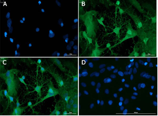
Immunofluorescence microscopy of Anti Doublecortin in PND1 cells. Post natal rat pup (PND1) heterogeneous brain cells were fixed with 4% PFA, permeabilization 0.3% Triton X 100. Incubated with 15ug/mL of anti Doublecortin primary antibody at 4C overnight. Counterstain Donkey anti Goat IgG DylightTM488 Conjugated antibody (p/n 605 741 125) at 5ug/mL for 1hr at room temperature. Nuclei were counterstained with DAPI present in mounting medium. Predicted cell localization is cytoplasm, cell projection. Image A). DAPI only. B). Anti Doublecortin Antibody + secondary. C). Merged A+B. D). Secondary only.
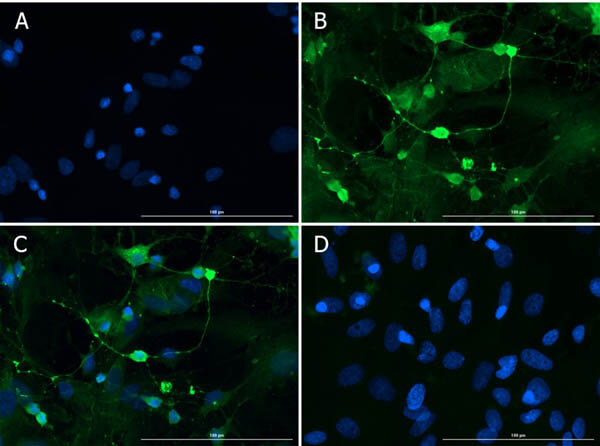
Immunofluorescence microscopy of Anti-Doublecortin in PND1 cells. Post-natal rat pup (PND1) heterogeneous brain cells were fixed with 4% PFA, permeabilization 0.3% Triton X-100. Incubated with 15ug/mL of anti-Doublecortin primary antibody at 4C overnight. Counterstain Donkey anti-Goat IgG DylightTM488 Conjugated antibody (p/n 605-741-125) at 5ug/mL for 1hr at room temperature. Nuclei were counterstained with DAPI present in mounting medium. Predicted cell localization is cytoplasm, cell projection. Note: localizes at neurite tips. Image A). DAPI only. B). Anti-Doublecortin Antibody + secondary. C). Merged A+B. D). Secondary only.
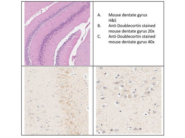
Immunohistochemistry of Goat Anti-Doublecortin antibody in mouse brain. Tissue: mouse dentate gyrus tissue. Antigen retrieval: heat induced (HIER) using Citrate Buffer for 20min. Primary antibody: Doublecortin antibody at 1:200 for 30min at RT. Secondary Antibody: Donkey Anti-Goat-HRP Antibody at 4μl/mL for 20min at RT. Counter Stain: Hematoxylin. Localization: Doublecortin (DCX) showed selective staining of soma and nuclei of cells in the dentate gyrus in mouse brain.
Western Blot of Goat Anti-Doublecortin Antibody. Lane 1: Opal Pre-stained MW ladder (p/n MB-210-0500). Lane 2: HEK293T Lysate (p/n W09-001-GX5) [15ug]. Lane 3: Rat brain, post natal, Whole Cell Lysate (p/n W12-001-MQ6) [15ug]. Lane 4: Rat pup PND2-6 - minimal cortex brain lysate [15ug]. Lane 5: Rat pup PND2-6 - minimal cortex brain lysate [35ug]. Primary antibody: Doublecortin antibody at 1:1000 for overnight at 4C. Secondary antibody: Donkey anti-goat secondary HRP antibody (p/n 605-703-125) at 1:40,000 for 1 hr at RT. Blocking: BlockOut (p/n MB-073) for 30 min at RT. Expected: ~40kDa. Observed size: ~45kDa.
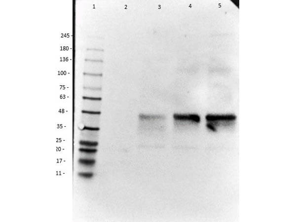
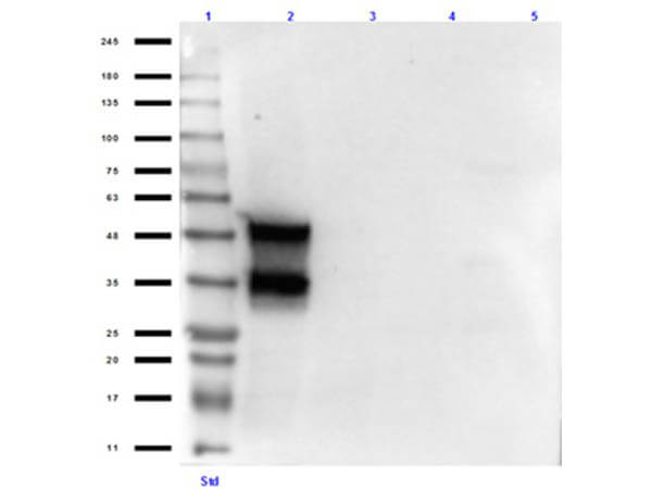
Western Blot of Goat Anti-Doublecortin Antibody. Lane 1: Opal Pre-stained MW ladder (p/n MB-210-0500). Lane 2: Doublecortin (human) Over Expressing HEK293T Lysate C-MYC/DDK tagged. Lane 3: HEK293T Lysate (p/n W09-001-GX5). Lane 4: Ms adult brain (normal tissue) (p/n W10-000-T004). Lane 5: Rat adult brain (normal tissue) (p/n W12-000-T077). Load: 10 ug per lane. Primary antibody: Doublecortin antibody at 1:1000 for overnight at 4C. Secondary antibody: Donkey anti-goat secondary HRP antibody (p/n 605-703-125) at 1:40,000 for 1 hr at RT. Blocking: BlockOut (p/n MB-073) for 30 min at RT. Expected: ~48kDa (overexpressing). Observed size: ~48/35kDa for Doublecortin.
|
|
|
|
Immunofluorescence microscopy of Anti Doublecortin in PND1 cells. Post natal rat pup (PND1) heterogeneous brain cells were fixed with 4% PFA, permeabilization 0.3% Triton X 100. Incubated with 15ug/mL of anti Doublecortin primary antibody at 4C overnight. Counterstain Donkey anti Goat IgG DylightTM488 Conjugated antibody (p/n 605 741 125) at 5ug/mL for 1hr at room temperature. Nuclei were counterstained with DAPI present in mounting medium. Predicted cell localization is cytoplasm, cell projection. Image A). DAPI only. B). Anti Doublecortin Antibody + secondary. C). Merged A+B. D). Secondary only.
|
|
|
| メーカー |
品番 |
包装 |
|
RKL
|
600-101-MH8
|
100 UG
|
※表示価格について
| 当社在庫 |
なし
|
| 納期目安 |
約10日
|
| 保存温度 |
-20℃
|
|

