|
※サムネイル画像をクリックすると拡大画像が表示されます。
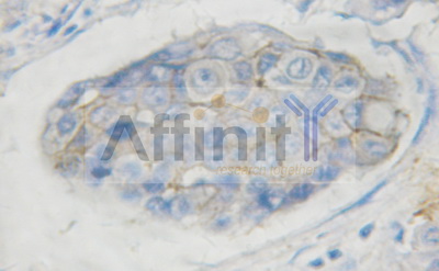
1/100 staining human breast carcinoma tissue by IHC-P. The sample was formaldehyde fixed and a heat mediated antigen retrieval step in citrate buffer was performed. The sample was then blocked and incubated with the antibody for 1.5 hours at 22ツーC. An HRP conjugated goat anti-rabbit antibody was used as the secondary antibody.
1:200 staining mouse testis tissue by IHC-P. The tissue was formaldehyde fixed and a heat mediated antigen retrieval step in citrate buffer was performed. The tissue was then blocked and incubated with the antibody for 1.5 hours at 22ツーC. An HRP conjugated goat anti-rabbit antibody was used as the secondary.

1:200 staining rat brain tissue by IHC-P. The tissue was formaldehyde fixed and a heat mediated antigen retrieval step in citrate buffer was performed. The tissue was then blocked and incubated with the antibody for 1.5 hours at 22ツーC. An HRP conjugated goat anti-rabbit antibody was used as the secondary.

1:200 staining rat kidney tissue by IHC-P. The tissue was formaldehyde fixed and a heat mediated antigen retrieval step in citrate buffer was performed. The tissue was then blocked and incubated with the antibody for 1.5 hours at 22ツーC. An HRP conjugated goat anti-rabbit antibody was used as the secondary.

1:200 staining rat kidney tissue by IHC-P. The tissue was formaldehyde fixed and a heat mediated antigen retrieval step in citrate buffer was performed. The tissue was then blocked and incubated with the antibody for 1.5 hours at 22ツーC. An HRP conjugated goat anti-rabbit antibody was used as the secondary.

1:200 staining rat lung tissue by IHC-P. The tissue was formaldehyde fixed and a heat mediated antigen retrieval step in citrate buffer was performed. The tissue was then blocked and incubated with the antibody for 1.5 hours at 22ツーC. An HRP conjugated goat anti-rabbit antibody was used as the secondary.

1:200 staining mouse testis tissue by IHC-P. The tissue was formaldehyde fixed and a heat mediated antigen retrieval step in citrate buffer was performed. The tissue was then blocked and incubated with the antibody for 1.5 hours at 22ツーC. An HRP conjugated goat anti-rabbit antibody was used as the secondary.

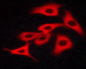
Staining H526 cells treated with SCF cells by IF/ICC. The samples were fixed with PFA and permeabilized in 0.1% saponin prior to blocking in 10% serum for 45 min at 37ツーC. The primary antibody was diluted 1/400 and incubated with the sample for 1 hour at 37ツーC. A Alexa Fluorツョ 594 conjugated goat polyclonal to rabbit IgG (H+L), diluted 1/600 was used as secondary antibody.
Western blot analysis of KIT phosphorylation expression in EGF treated HepG2 whole cells lysates. The lane on the left is treated with the antigen-specific peptide.

Western blot analysis of Phospho-KIT (Tyr703) expression in various lysates

|
|
|
|
1/100 staining human breast carcinoma tissue by IHC-P. The sample was formaldehyde fixed and a heat mediated antigen retrieval step in citrate buffer was performed. The sample was then blocked and incubated with the antibody for 1.5 hours at 22ツーC. An HRP conjugated goat anti-rabbit antibody was used as the secondary antibody.
|
|
| 別品名 |
KIT, C-Kit, CD117, PBT, Piebald trait, Piebald trait protein, Proto-oncogene c-Kit, SCFR, Soluble KIT variant 1, Tyrosine-protein kinase Kit, p145 c-kit, CD117 antigen
|
| 種由来 |
Human
|
| 交差種 |
Human
Mouse
Rat
|
| 適用 |
Western Blot
Enzyme Linked Immunosorbent Assay
Immunohistochemistry
Immuno Fluorescence
|
| 免疫動物 |
Rabbit
|
| 抗体クラス |
IgG
|
| 標識物 |
Unlabeled
|
| 精製度 |
Affinity Purified
|
| 翻訳後修飾 |
リン酸化
|
| GENE ID |
3815
|
| Gene Symbol |
KIT
|
|
| メーカー |
品番 |
包装 |
|
LSP
|
LS-C801010-100
|
100 UL
|
※表示価格について
| 当社在庫 |
なし
|
| 納期目安 |
約1ヶ月
|
| 保存温度 |
-20℃
|
|
※当社では商品情報の適切な管理に努めておりますが、表示される法規制情報は最新でない可能性があります。
また法規制情報の表示が無いものは、必ずしも法規制に非該当であることを示すものではありません。
商品のお届け前に最新の製品法規制情報をお求めの際はこちらへお問い合わせください。
|
※当社取り扱いの試薬・機器製品および受託サービス・創薬支援サービス(納品物、解析データ等)は、研究用としてのみ販売しております。
人や動物の医療用・臨床診断用・食品用としては、使用しないように、十分ご注意ください。
法規制欄に体外診断用医薬品と記載のものは除きます。
|
|
※リンク先での文献等のダウンロードに際しましては、掲載元の規約遵守をお願いします。
|
|
※CAS Registry Numbers have not been verified by CAS and may be inaccurate.
|
