|
※サムネイル画像をクリックすると拡大画像が表示されます。
Immunohistochemical staining of paraffin-embedded human melanoma using anti-FOS clone UMAB221 at 1:200 dilution of 0.5 mg/mL and detection with Polink2 Broad HRP DAB.requires heat-induced epitope retrieval with ACCEL. (pH8.7) biocare pressure cooker. The image shows nuclear staining in tumor cells.

Immunohistochemical staining of paraffin-embedded human pancreas tissue within the normal limits using anti-FOS mouse monoclonal antibody. HIER pretreatment was done with 1mM EDTA in 10mM Tris buffer. (pH8.0) at 120ツーC for 2.5 minutes.was diluted 1:200 and detection was done with HRP secondary and DAB chromogen. Here we see nuclear staining.
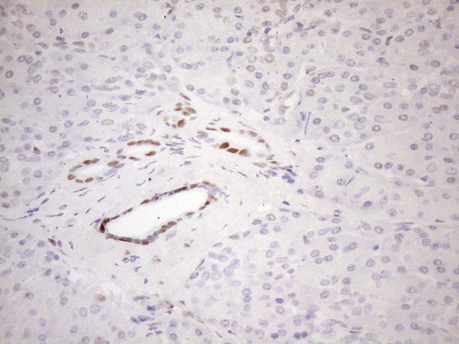
Immunohistochemical staining of paraffin-embedded Carcinoma of Human pancreas tissue using anti-FOS mouse monoclonal antibody.HIER pretreatment was done with 1mM EDTA in 10mM Tris buffer. (pH8.0) at 120ツーC for 2.5 minutes.was diluted 1:200 and detection was done with HRP secondary and DAB chromogen. Nuclear staining seen in tumor cells.
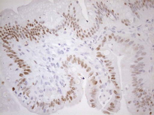
Immunohistochemical staining of paraffin-embedded carcinoma of human thyroid tissue using anti-FOS mouse monoclonal antibody.HIER pretreatment was done with 1mM EDTA in 10mM Tris buffer. (pH8.0) at 120ツーC for 2.5 minutes.was diluted 1:200 and detection was done with HRP secondary and DAB chromogen. Nuclear staining seen in tumor cells.
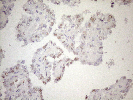
Immunohistochemical staining of paraffin-embedded Carcinoma of Human prostate tissue using anti-FOS mouse monoclonal antibody.HIER pretreatment was done with 1mM EDTA in 10mM Tris buffer. (pH8.0) at 120ツーC for 2.5 minutes.was diluted 1:200 and detection was done with HRP secondary and DAB chromogen. Nuclear staining seen in tumor cells.
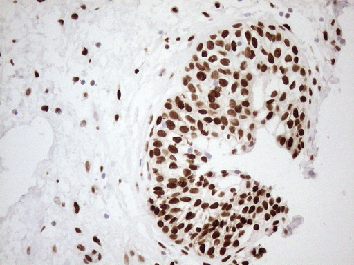
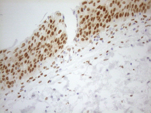
Immunohistochemical staining of paraffin-embedded human bladder tissue within the normal limits using anti-FOS mouse monoclonal antibody.HIER pretreatment was done with 1mM EDTA in 10mM Tris buffer. (pH8.0) at 120ツーC for 2.5 minutes.was diluted 1:200 and detection was done with HRP secondary and DAB chromogen. Nuclear staining seen in epithelial cells.
OriGene overexpression protein microarray chip was immunostained with UltraMAB anti-FOS mouse monoclonal antibody. The positive reactive proteins are highlighted with two red arrows in the enlarged subarray. All the positive controls spotted in this subarray are also labeled for clarification. (1:100)

|
|
|
|
Immunohistochemical staining of paraffin-embedded human melanoma using anti-FOS clone UMAB221 at 1:200 dilution of 0.5 mg/mL and detection with Polink2 Broad HRP DAB.requires heat-induced epitope retrieval with ACCEL. (pH8.7) biocare pressure cooker. The image shows nuclear staining in tumor cells.
|
|
| 別品名 |
FOS, Activator protein 1, AP-1, Cellular oncogene c-fos, Cellular oncogene fos, Proto-oncogene c-Fos, v-fos, C-FOS, G0s7, p55
|
| 種由来 |
Human
|
| 交差種 |
Human
|
| 適用 |
Western Blot
Immunohistochemistry
|
| 免疫動物 |
Mouse
|
| クローン |
UMAB221
|
| 抗体クラス |
IgG1
|
| 標識物 |
Unlabeled
|
| 精製度 |
Affinity Purified
|
| GENE ID |
2353
|
| Gene Symbol |
FOS
|
| 性状 |
Carrier Free
|
|
| メーカー |
品番 |
包装 |
|
LSP
|
LS-C796992-100
|
100 UG
|
※表示価格について
| 当社在庫 |
なし
|
| 納期目安 |
約1ヶ月
|
| 保存温度 |
-20℃
|
|
※当社では商品情報の適切な管理に努めておりますが、表示される法規制情報は最新でない可能性があります。
また法規制情報の表示が無いものは、必ずしも法規制に非該当であることを示すものではありません。
商品のお届け前に最新の製品法規制情報をお求めの際はこちらへお問い合わせください。
|
※当社取り扱いの試薬・機器製品および受託サービス・創薬支援サービス(納品物、解析データ等)は、研究用としてのみ販売しております。
人や動物の医療用・臨床診断用・食品用としては、使用しないように、十分ご注意ください。
法規制欄に体外診断用医薬品と記載のものは除きます。
|
|
※リンク先での文献等のダウンロードに際しましては、掲載元の規約遵守をお願いします。
|
|
※CAS Registry Numbers have not been verified by CAS and may be inaccurate.
|


