

| 種由来 |
Mouse
|
| 標識物 |
Fluorescein Isothiocyanate
|
| 励起波長 |
492
|
| 蛍光波長 |
518
|
| 精製度 |
Affinity Purified
|
| 適用 |
Immunohistochemistry
Flow Cytometry
Fluorescence based plate assays
|
| 免疫動物 |
Goat
|
| 抗体クラス |
IgG
|
| 交差種 |
Mouse
|
| 非交差(吸収処理)種 |
Human
|
| GENE ID |
16017
|
| Accession No.(Gene/Protein) |
P01868 P01869
|
| 形状 |
液状
|
| 使用目的 |
Goat Anti-Mouse IgG1-FITC antibody with minimal reactivity to human proteins for use in flow cytometry, immunohistochemistry / immunocytochemistry, and western blotting assays.
|
|
※サムネイル画像をクリックすると拡大画像が表示されます。
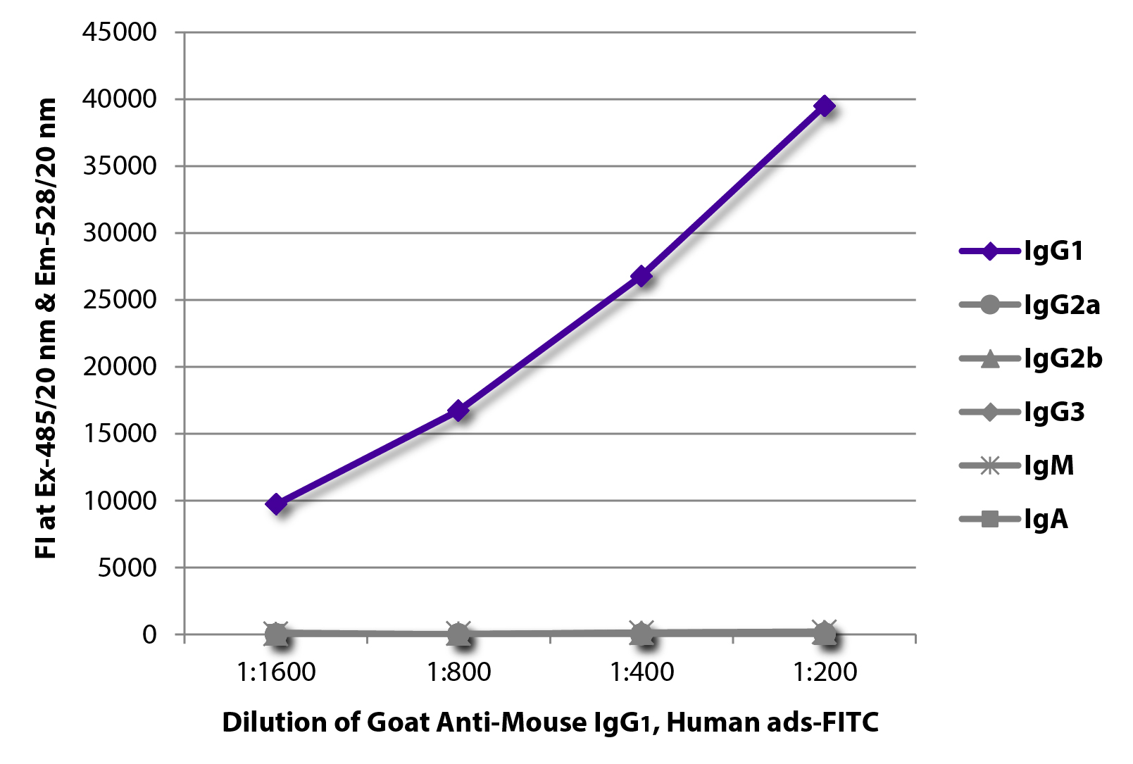
FLISA plate was coated with purified mouse IgG1, IgG2a, IgG2b, IgG3, IgM, and IgA. Immunoglobulins were detected with serially diluted Goat Anti-Mouse IgG1, Human ads-FITC (SB Cat. No. 1070-02).
Zebrafish embryos were stained with anti-atrial myosin heavy chain and anti-sacromeric myosin heavy chain followed by Goat Anti-Mouse IgG1, Human ads-FITC (SB Cat. No. 1070-02) and Goat Anti-Mouse IgG2b, Human ads-TRITC (SB Cat. No. 1090-03) secondary antibodies.Image from Zhu D, Fang Y, Gao K, Shen J, Zhong TP, Li F. Vegfa impacts early myocardium development in zebrafish. Int J Mol Sci. 2017;18:444. Figure 4(g)
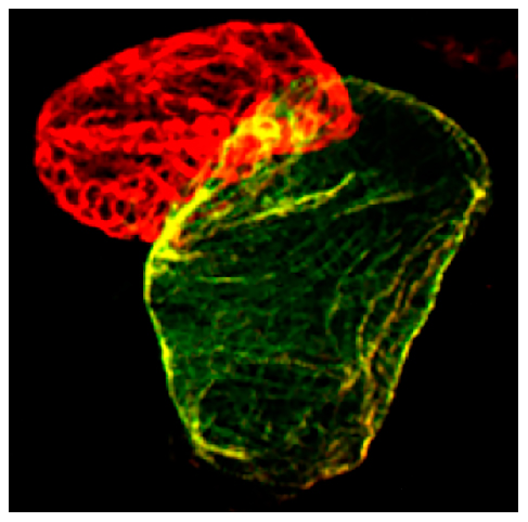
Tg(myl7:nDSRed) transgenic zebrafish embryos were stained with anti-atrial myosin heavy chain and anti-DsRed followed by anti-rabbit AF594 and Goat Anti-Mouse IgG1, Human ads-FITC (SB Cat. No. 1070-02) secondary antibodies.Image from Zhu D, Fang Y, Gao K, Shen J, Zhong TP, Li F. Vegfa impacts early myocardium development in zebrafish. Int J Mol Sci. 2017;18:444. Figure 5(b)

Frozen equine sacroid tissue section was stained with anti-CD4 followed by Goat Anti-Mouse IgG1, Human ads-FITC (SB Cat. No. 1070-02) and DAPI.Image from Wilson AD, Hicks C. Both tumour cells and infiltrating T-cells in equine sarcoids express FOXP3 associated with an immune-supressed cytokine microenvironment. Vet Res. 2016;47:55. Figure 2(h)

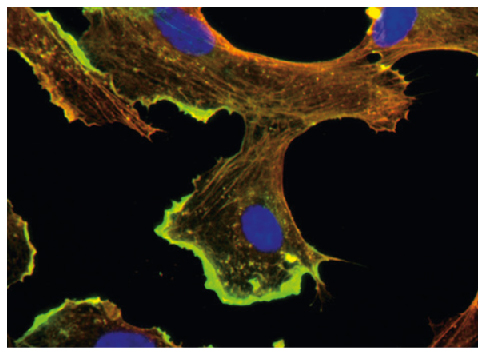
BMH29L cells were stained with anti-β-actin and anti-γ-actin followed by Goat Anti-Mouse IgG1, Human ads-FITC (SB Cat. No. 1070-02) and Goat Anti-Mouse IgG2b, Human ads-TRITC (SB Cat. No. 1090-03) and DAPI.Image from Pasquier E, Tuset M, Sinnappan S, Carnell M, Macmillan A, Kavallaris M. γ-Actin plays a key role in endothelial cell motility and neovessel maintenance. Vasc Cell. 2015;7:2. BMC Med Genet. 2017;18:44. Figure 1
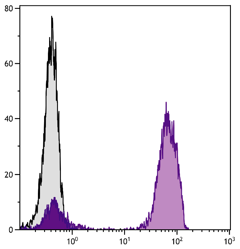
Human peripheral blood lymphocytes were stained with Mouse Anti-Human CD3-UNLB followed by Goat Anti-Mouse IgG1, Human ads-FITC (SB Cat. No. 1070-02).
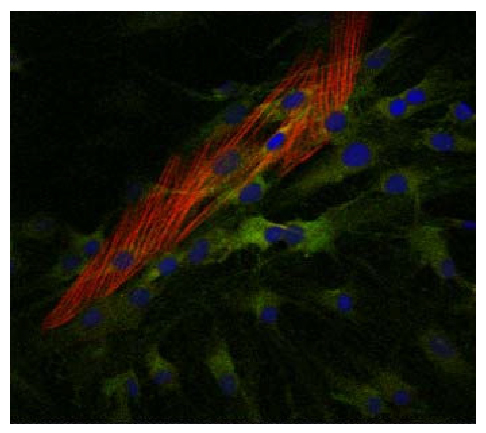
Human myofibroblasts were stained with anti-COX2 and anti-α-SMA followed by Goat Anti-Mouse IgG2a, Human ads-TXRD (SB Cat. No. 1080-07), Goat Anti-Mouse IgG1, Human ads-FITC (SB Cat. No. 1070-02), and DAPI.Image from Mattyasovszky SG, Hofmann A, Brochhausen C, Ritz U, Kuhn S, Wollstadter J, et al. The effect of the pro-inflammatory cytokine tumor necrosis factor-alpha on human joint capsule myofibroblasts. Arthritis Res Ther. 2010;12:R4. Figure 7(a)
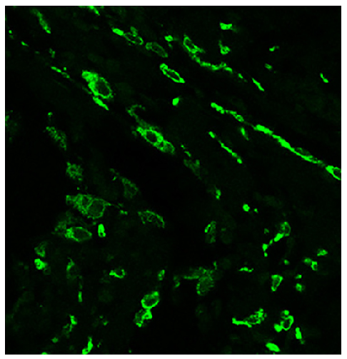
Paraffin embedded human supraspinatus tendon tear section was stained with anti-CD206 followed by Goat Anti-Mouse IgG1, Human ads-FITC (SB Cat. No. 1070-02).Image from Goodier HC, Carr AJ, Snelling SJ, Roche L, Wheway K, Watkins B, et al. Comparison of transforming growth factor beta expression in healthy and diseased human tendon. Arthritis Res Ther. 2016;18:48. Figure 5
Frozen newborn mouse cartilage section was stained with Mouse IgG1-UNLB isotype control (SB Cat. No. 0102-01; left) and Mouse Anti-Type II Collagen-UNLB (right) followed by Goat Anti-Mouse IgG1, Human ads-FITC (SB Cat. No. 1070-02) and DAPI.

|
|
|
|
FLISA plate was coated with purified mouse IgG1, IgG2a, IgG2b, IgG3, IgM, and IgA. Immunoglobulins were detected with serially diluted Goat Anti-Mouse IgG1, Human ads-FITC (SB Cat. No. 1070-02).
|
|
|
| メーカー |
品番 |
包装 |
|
SBA
|
1070-02
|
1 MG
[1.0 mg/mL]
|
※表示価格について
|

