|
※サムネイル画像をクリックすると拡大画像が表示されます。
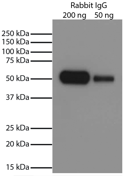
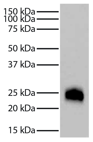
Rabbit IgG-UNLB (SB Cat. No. 0111-01) was resolved by electrophoresis, transferred to PVDF membrane, and visualized using Goat Anti-Rabbit IgG-HRP (SB Cat. No. 4030-05) secondary antibody and chemiluminescent detection.
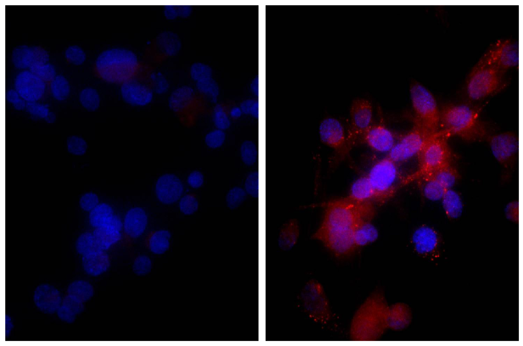
Human hepatocellular carcinoma cell line Hep G2 was stained with Rabbit IgG-UNLB isotype control (SB Cat. No. 0111-01; left) and Rabbit Anti-Human DR5-UNLB (SB Cat. No. 6600-01; right) followed by Donkey Anti-Rabbit IgG(H+L), Mouse/Rat/Human SP ads-BIOT (SB Cat. No. 6440-08, Streptavidin-CY3 (SB Cat. No. 7100-12), and DAPI.
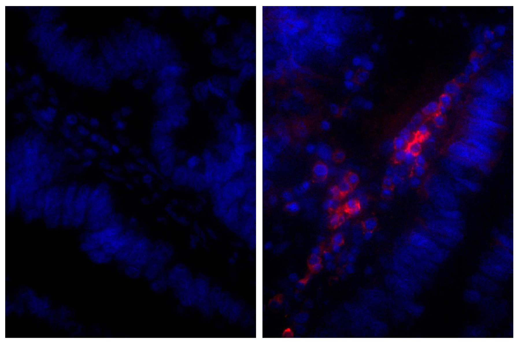
Paraffin embedded human gastric cancer tissue was stained with Rabbit IgG-UNLB isotype control (SB Cat. No. 0111-01; left) and Rabbit Anti-Human IgG(H+L), Mouse ads-UNLB (SB Cat. No. 6145-01; right) followed by Donkey Anti-Rabbit IgG(H+L), Mouse/Rat/Human SP ads-AF555 (SB Cat. No. 6440-32) and DAPI.
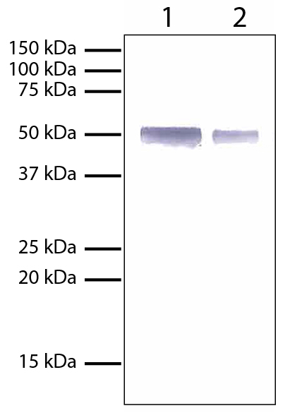
Lane 1 - 1 mg Rabbit IgGLane 2 - 0.5 mg Rabbit IgGRabbit IgG (SB Cat. No. 0111-01) was resolved by electrophoresis under reducing conditions, transferred to PVDF membrane, and probed with Mouse Anti-Rabbit IgG-AP (SB Cat. No. 4090-04). Proteins were visualized using BCIP/NBT One Component Membrane Substrate (Blue), Solution (SB Cat. No. 0430-01).
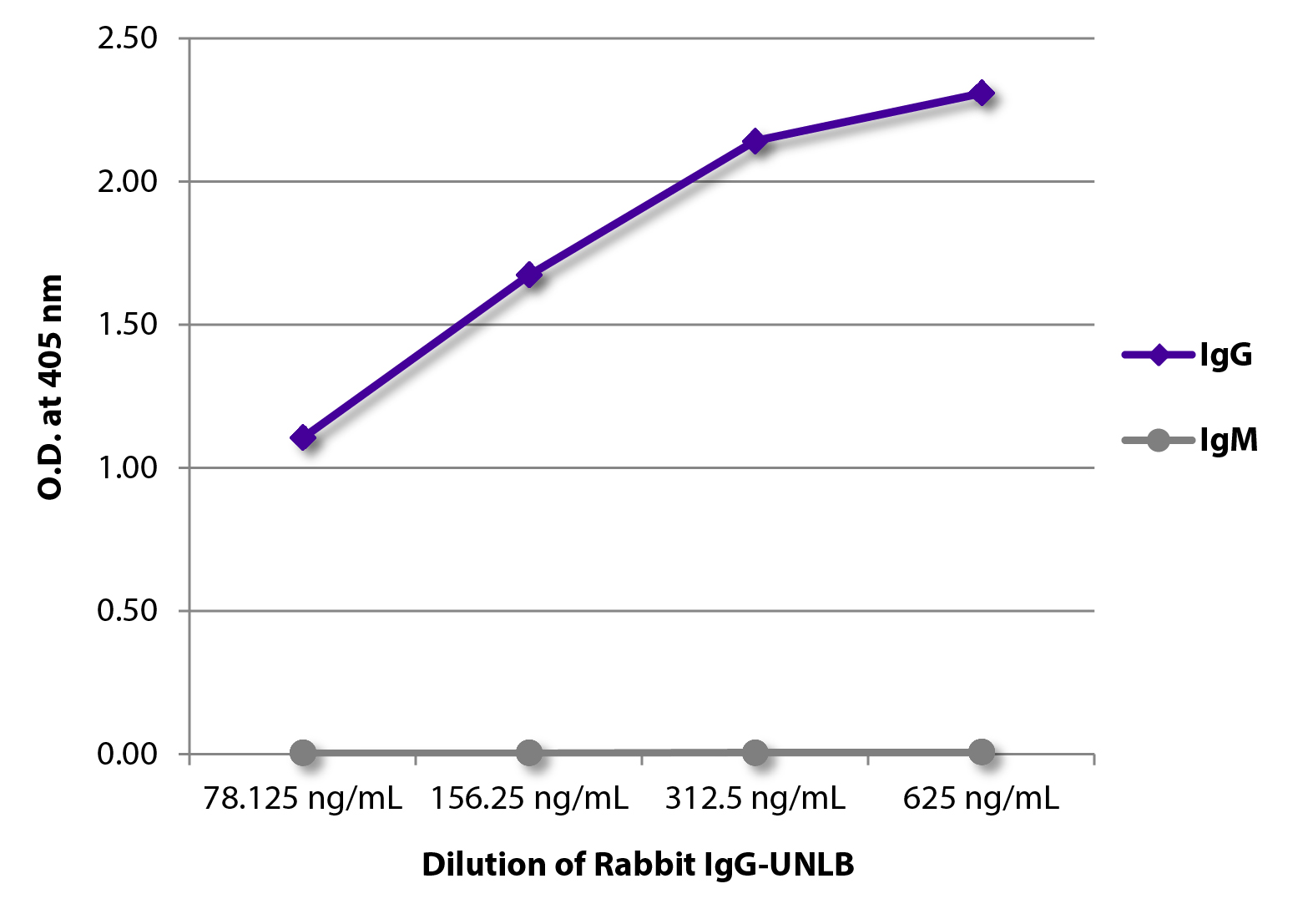
ELISA plate was coated with serially diluted Rabbit IgG-UNLB (SB Cat. No. 0111-01). Immunoglobulin was detected with Goat Anti-Rabbit IgG-BIOT (SB Cat. No. 4030-08) and Goat Anti-Rabbit IgM-BIOT (SB Cat. No. 4020-08) followed by Streptavidin-HRP (SB Cat No. 7100-05) and quantified.
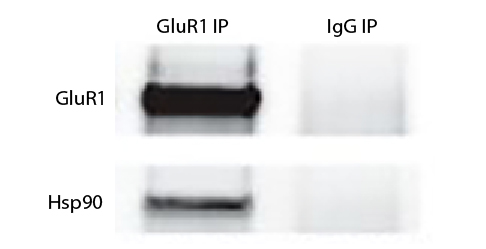
Tissue lysate from rTgFKBP5 double transgenic mouse hippocampus was pre-cleared with Mouse IgG-UNLB (SB Cat. No. 0107-01) and immunoprecipitated with anti-GluR1 or Rabbit IgG-UNLB (SB Cat. No. 0111-01). Protein was resolved by electrophoresis, transferred to membrane, and probed with anti-GluR1 or anti-HSP90 followed by Goat Anti-Rabbit IgG(H+L), Mouse/Human ads-BIOT (SB Cat. No. 4050-08), Goat Anti-Mouse IgG, Human ads-HRP (SB Cat. No. 1030-05), an HRP conjugated biotin binding protein, and chemiluminescent detection.Image from Blair LJ, Criado-Marrero M, Zheng D, Wang X, Kamath S, Nordhues BA, et al. The disease-associated chaperone FKBP51 impairs cognitive function by accelerating AMPA receptor recycling. eNeuro. 2019;6:e0242-18.2019. Figure 8(c)
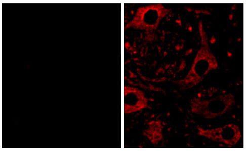
Frozen mouse spinal cord sections were stained with anti-BAFF-R (right) and Rabbit IgG-UNLB (SB Cat No. 0111-01; left) followed by a CY5 conjugated secondary antibody.Images from Tada S, Yasui T, Nakatsuji Y, Okuno T, Koda T, Mochizuki H, et al. BAFF controls neural cell survival through BAFF receptor. PLoS One. 2013;8(7):e70924. Figure 1(c/d)
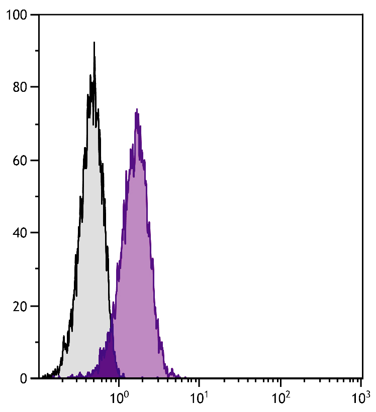
Human T cell leukemia cell line Jurkat was stained with Rabbit IgG-UNLB (SB Cat. No. 0111-01; gray) and Rabbit Anti-Human DR5-UNLB (SB Cat. No. 6600-01) followed by Mouse Anti-Rabbit IgG-PE (SB Cat. No. 4090-09).
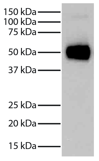
Rabbit IgG-UNLB (SB Cat. No. 0111-01) was resolved by electrophoresis under reducing conditions, transferred to PVDF membrane, and probed with Mouse Anti-Rabbit Light Chain-HRP (SB Cat. No. 4060-05) followed by chemiluminescent detection.
Rabbit IgG-UNLB (SB Cat. No. 0111-01) was resolved by electrophoresis under reducing conditions, transferred to PVDF membrane, and probed with Goat Anti-Rabbit IgG-BIOT (SB Cat. No. 4030-08) followed by Streptavidin-HRP (SB Cat. No. 7105-05) and chemiluminescent detection.
|
|
|
|
Rabbit IgG-UNLB (SB Cat. No. 0111-01) was resolved by electrophoresis, transferred to PVDF membrane, and visualized using Goat Anti-Rabbit IgG-HRP (SB Cat. No. 4030-05) secondary antibody and chemiluminescent detection.
|
|
| 種由来 |
Rabbit
|
| 由来詳細 |
Normal rabbit serum
|
| 適用 |
Enzyme Linked Immunosorbent Assay
Immunocytochemistry (cell)
Flow Cytometry
|
| 抗体クラス |
IgG
|
| 標識物 |
Unlabeled
|
| 発現系 |
Native
|
| 使用目的 |
Purified Rabbit IgG for use as a control in flow cytometry, immunocytochemistry / immunocytochemistry, western blotting, immunoprecipitation, ChIP, microarray, and in vitro assays and as a standard in ELISA.
|
| 構成内容 |
[Buffer Formulation]Borate buffered saline, pH 8.2
|
|
| メーカー |
品番 |
包装 |
|
SBA
|
0111-01
|
10 MG
[5 mg/mL]
|
※表示価格について
|
※当社では商品情報の適切な管理に努めておりますが、表示される法規制情報は最新でない可能性があります。
また法規制情報の表示が無いものは、必ずしも法規制に非該当であることを示すものではありません。
商品のお届け前に最新の製品法規制情報をお求めの際はこちらへお問い合わせください。
|
※当社取り扱いの試薬・機器製品および受託サービス・創薬支援サービス(納品物、解析データ等)は、研究用としてのみ販売しております。
人や動物の医療用・臨床診断用・食品用としては、使用しないように、十分ご注意ください。
法規制欄に体外診断用医薬品と記載のものは除きます。
|
|
※リンク先での文献等のダウンロードに際しましては、掲載元の規約遵守をお願いします。
|
|
※CAS Registry Numbers have not been verified by CAS and may be inaccurate.
|
