|
※サムネイル画像をクリックすると拡大画像が表示されます。
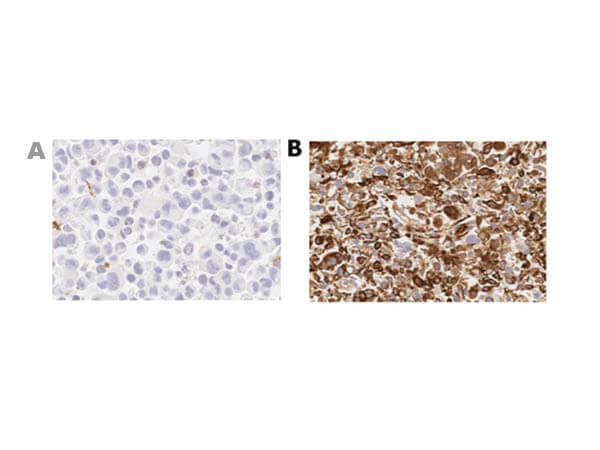
Immunohistochemistry of Rabbit Anti-MUC4 Antibody. Tissue : A) Negative control PANC1; B) Positive control HPAC. Primary Antibody: Anti-MUC4 at 1:1000. Secondary Antibody: Ready-to-Use Anti-Rabbit. Staining: DAB. Counter Stain: Hematoxylin.
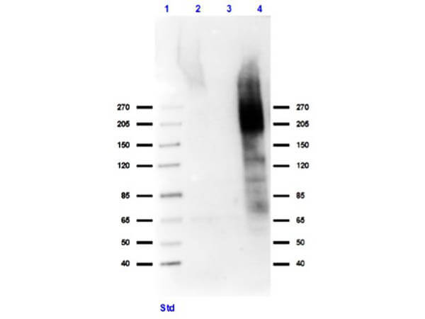
Western Blot of Rabbit Anti-MUC4 Antibody. Lane 1: Spectra Multicolor Ladder. Lane 2: MDA-MB-231 lysate (p/n W09-001-GK6). Lane 3: HeLa lysate (p/n W09-001-364). Lane 4: HPAC Lysate (MUC4+) positive control. Primary Antibody: Anti-MUC4 at 1:1000 in (p/n MB-073) at RT for 30 min. Secondary Antibody: Goat Anti-Rabbit IgG HRP (p/n 611-103-122) at 1:70,000 in MB-073 for 1 hr at RT. Expect: ~550-930 kDa. Observed: ~200-300 kDa. Highly glycosylated protein.
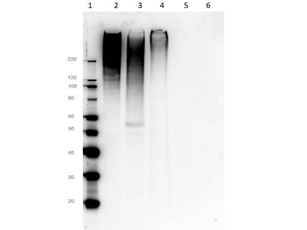
Western Blot of Rabbit Anti-MUC4 Antibody. Lane 1: Spectra Multicolor Ladder. Lane 2: HPAF-II [MUC4+]. Lane 3: HPAC [MUC4+]. Lane 4: Capan-2 [MUC4+]. Lane 5: MiaPaCa2 [MUC4-]. Lane 6: Panc-1 (p/n W09-001-GM2) [MUC4-]. Primary Antibody: Anti-MUC4 at 1:1000 in 2% BSA at RT for 2 hrs. Secondary Antibody: Goat Anti-Rabbit IgG HRP (p/n 611-103-122) at 1:70,000 in 5% BSA/PBS/0.1 Tween 20 for 1 hr at RT. Expect: ~550-930 kDa. Observed: ~200-300 kDa. Highly glycosylated protein.
|
|
|
|
Immunohistochemistry of Rabbit Anti-MUC4 Antibody. Tissue : A) Negative control PANC1; B) Positive control HPAC. Primary Antibody: Anti-MUC4 at 1:1000. Secondary Antibody: Ready-to-Use Anti-Rabbit. Staining: DAB. Counter Stain: Hematoxylin.
|
|
| 別品名 |
Rabbit Anti-MUC4 Antibody, Rabbit Anti-Mucin 4 Antibody, MUC4, Mucin-4, Ascites sialoglycoprotein, ASGP, Pancreatic adenocarcinoma mucin, Testis mucin, Tracheobronchial mucin, Mucin-4 alpha chain, Ascites sialoglycoprotein 1, ASGP-1, Mucin-4 beta chain, Ascites sialoglycoprotein 2, ASGP-2, MUC
|
| 交差種 |
Human
|
| 適用 |
Western Blot
Enzyme Linked Immunosorbent Assay
Immunohistochemistry
|
| 免疫動物 |
Rabbit
|
| 標識物 |
Unlabeled
|
| GENE ID |
4585
|
| Accession No.(Gene/Protein) |
NP_004523.3, Q99102
|
| Gene Symbol |
MUC4
|
| 参考文献 |
[Pub Med ID]30952968
|
|
| メーカー |
品番 |
包装 |
|
RKL
|
600-401-GY2
|
100 UG
|
※表示価格について
| 当社在庫 |
なし
|
| 納期目安 |
約10日
|
| 保存温度 |
-20℃
|
|
※当社では商品情報の適切な管理に努めておりますが、表示される法規制情報は最新でない可能性があります。
また法規制情報の表示が無いものは、必ずしも法規制に非該当であることを示すものではありません。
商品のお届け前に最新の製品法規制情報をお求めの際はこちらへお問い合わせください。
|
※当社取り扱いの試薬・機器製品および受託サービス・創薬支援サービス(納品物、解析データ等)は、研究用としてのみ販売しております。
人や動物の医療用・臨床診断用・食品用としては、使用しないように、十分ご注意ください。
法規制欄に体外診断用医薬品と記載のものは除きます。
|
|
※リンク先での文献等のダウンロードに際しましては、掲載元の規約遵守をお願いします。
|
|
※CAS Registry Numbers have not been verified by CAS and may be inaccurate.
|
