|
※サムネイル画像をクリックすると拡大画像が表示されます。
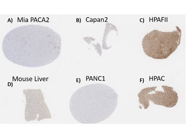
Immunohistochemistry of Mouse Anti-MUC4 Antibody. Tissue/Cell: A) hu Mia PACA2 [-]. B) hu Capan2 pancreatic ductal adenocarcinoma cell line [+]. C) hu HPAFII cell pellet [+]. D) Control mouse liver [-]. E) hu Control PANC1 cell pellet [-]. F) hu HPAC cell pellet [+]. Tissues: FFPE containing normal and malignant pancreatic tissues. Cell pellets: Appr. 10 million cells were collected from flasks without enzymatic treatment and centrifuged; thrombin and Fibrinogen was added. The resulting pellet of clotted cells was collected. Fixative: 10% neutral buffered formalin at RT. Antigen Retrieval: HIER citrate buffer for 20 min. Primary Antibody: Anti-MUC4 Antibody at 1:1000 in A, B, C, F. Isotype control reagent used in negative control D, E. Secondary Antibody: Anti-Mouse IgG. Counter Stain: Bond Polymer Refine Detection Kit. Analysis Results: No staining was observed for control liver and PANC1 (MUC4−) cell pellets while HPAFII and HPAC pellets that are known to express large amounts of MUC4 were strongly and diffusely positive. Some non-specific, acellular staining is seen within Mia PACA2 (MUC4−) cells.
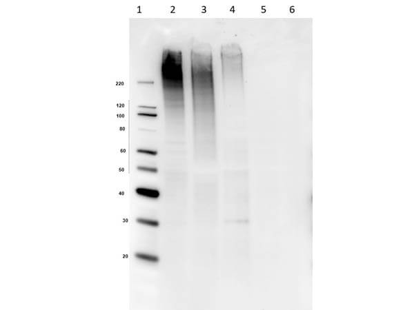
Western Blot of Mouse Anti-MUC4 Antibody. Lane 1: Spectra Multicolor Ladder. Lane 2: HPAF-II (MUC4+). Lane 3: HPAC (MUC4+). Lane 4: Capan-2 (MUC4+). Lane 5: MiaPaCa2 (MUC4-). Lane 6: Panc-1 (MUC4-). Primary Antibody: Anti-MUC4 at 1nM in 2% BSA at RT for 2 hrs. Secondary Antibody: Goat Anti-Mouse IgG HRP (610-103-121) at 1:70,000 in 5% BSA/PBS/0.1 Tween 20 for 1 hr at RT. Expect: ~550-930 kDa. Observed: ~200-300 kDa. Highly glycosylated protein.
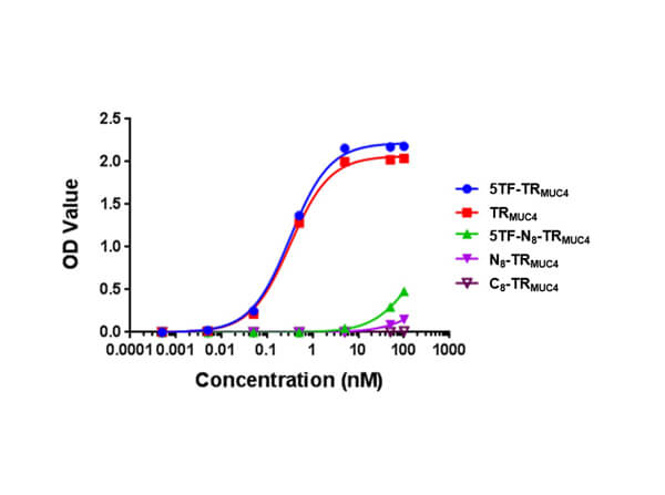
ELISA results from BSA constructs (0.3μg/mL) coated on plates and screened using the following antisera: anti-5TFag-TRMUC4 serum (1:300), affinity pure anti-5TFag-TRMUC4 polyclonal antibody (15μg/mL) and unimmunized rabbit serum (1:300). Primary antisera were added to well plates and serially diluted by half Log10 dilutions. Assay performed using Blocking buffer (3 % BSA), alkaline phosphatase conjugated secondary antibodies, and PNPP (10 mg) in pH 9.8 DEA buffer.
|
|
|
|
Immunohistochemistry of Mouse Anti-MUC4 Antibody. Tissue/Cell: A) hu Mia PACA2 [-]. B) hu Capan2 pancreatic ductal adenocarcinoma cell line [+]. C) hu HPAFII cell pellet [+]. D) Control mouse liver [-]. E) hu Control PANC1 cell pellet [-]. F) hu HPAC cell pellet [+]. Tissues: FFPE containing normal and malignant pancreatic tissues. Cell pellets: Appr. 10 million cells were collected from flasks without enzymatic treatment and centrifuged; thrombin and Fibrinogen was added. The resulting pellet of clotted cells was collected. Fixative: 10% neutral buffered formalin at RT. Antigen Retrieval: HIER citrate buffer for 20 min. Primary Antibody: Anti-MUC4 Antibody at 1:1000 in A, B, C, F. Isotype control reagent used in negative control D, E. Secondary Antibody: Anti-Mouse IgG. Counter Stain: Bond Polymer Refine Detection Kit. Analysis Results: No staining was observed for control liver and PANC1 (MUC4−) cell pellets while HPAFII and HPAC pellets that are known to express large amounts of MUC4 were strongly and diffusely positive. Some non-specific, acellular staining is seen within Mia PACA2 (MUC4−) cells.
|
|
| 別品名 |
Mouse Anti-MUC4 Antibody, Mouse Anti-Mucin 4 Antibody, MUC4, Mucin-4, Ascites sialoglycoprotein, ASGP, Pancreatic adenocarcinoma mucin, Testis mucin, Tracheobronchial mucin, Mucin-4 alpha chain, Ascites sialoglycoprotein 1, ASGP-1, Mucin-4 beta chain, Ascites sialoglycoprotein 2, ASGP-2, MUC
|
| 交差種 |
Human
|
| 適用 |
Western Blot
Enzyme Linked Immunosorbent Assay
Immunohistochemistry
|
| 免疫動物 |
Mouse
|
| クローン |
3C7.D8.E11.F5
|
| 標識物 |
Unlabeled
|
| GENE ID |
4585
|
| Accession No.(Gene/Protein) |
NP_004523.3, Q99102
|
| Gene Symbol |
MUC4
|
| 参考文献 |
[Pub Med ID]30952968
|
|
| メーカー |
品番 |
包装 |
|
RKL
|
200-301-GY2
|
100 UG
|
※表示価格について
| 当社在庫 |
なし
|
| 納期目安 |
約10日
|
| 保存温度 |
-20℃
|
|
※当社では商品情報の適切な管理に努めておりますが、表示される法規制情報は最新でない可能性があります。
また法規制情報の表示が無いものは、必ずしも法規制に非該当であることを示すものではありません。
商品のお届け前に最新の製品法規制情報をお求めの際はこちらへお問い合わせください。
|
※当社取り扱いの試薬・機器製品および受託サービス・創薬支援サービス(納品物、解析データ等)は、研究用としてのみ販売しております。
人や動物の医療用・臨床診断用・食品用としては、使用しないように、十分ご注意ください。
法規制欄に体外診断用医薬品と記載のものは除きます。
|
|
※リンク先での文献等のダウンロードに際しましては、掲載元の規約遵守をお願いします。
|
|
※CAS Registry Numbers have not been verified by CAS and may be inaccurate.
|
