|
※サムネイル画像をクリックすると拡大画像が表示されます。
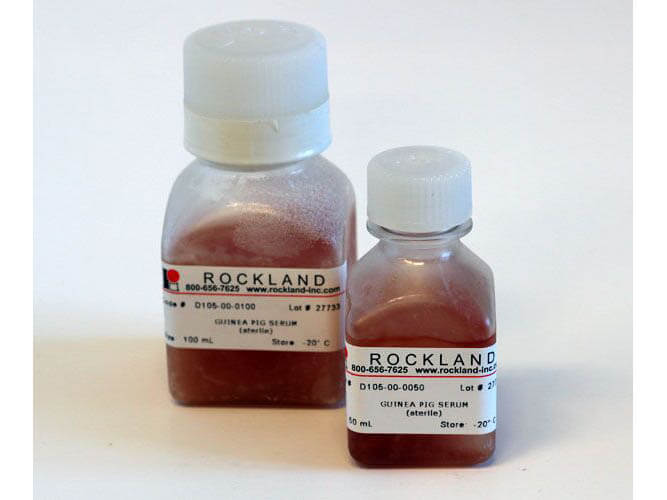
Guinea Pig Serum is used as a supplement to cell culture media. Guinea Pig Serum provides a broad spectrum of macromolecules, carrier proteins for lipoid substances and trace elements, attachment and spreading factors, low molecular weight nutrients, and hormones and growth factors that promote cell growth and health. Be certain to maintain Good Cell Culture Practice, and maintain sterility of cultures that require media supplementation.
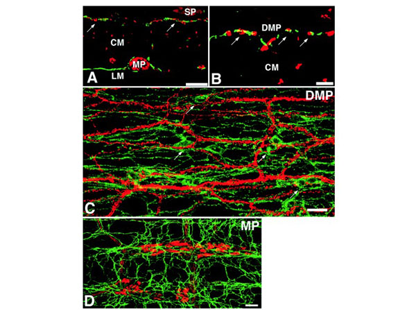
Double-labeling immunofluorescence for c-Kit and nitric oxide synthase (NOS) in the guinea-pig small intestine. The immunofluorescence for FITC (green) and Texas Red (red) reveals c-Kit-like immunoreactivity (c-Kit-LI) and NOS-like immunoreactivity (NOS-LI), respectively. Secondary Antibodies blocked with 2% guinea-pig serum (p/n D105-00-0050) in 1% BSA-PBS.Figs. A and B were obtained from cross sections of muscle fibers of the circular muscle layer, while Figs. C and D were from whole mount preparations. DMP: deep muscular plexus, MP: myenteric plexus, SP: submucosal plexus, CM: circular muscle layer, LM: longitudinal muscle layer. (A) The overlapping of c-Kit-LI and NOS-LI is more prominent in the DMP arrows than in the myenteric plexus. The dense labeling of NOS-LI is also observed in the SP and myenteric plexus but is absent in the LM. Scale bars = 50 mm. (B) A higher magnification of the DMP. The yellow labeling indicates the close association between c-Kit-LI and NOS-LI (arrows). Scale bar = 10 mm. (C) In the DMP, c-Kit-LI exhibits several long and fine processes which constitute a network. NOS-LI bifurcates and fuses the neighboring lines, forming a dense network running parallel to the long axis of muscle fibers of the CM (in the horizontal direction of the figure). The dots or strands of the labeling with NOS-LI enclose c-Kit-LI (arrows). Scale bar = 20 mm. (D) In the myenteric plexus, NOS-LI is detected within the ganglion strands or connecting strands. In the ganglion strands, NOS-LI exhibits several short and flattened processes as well as a long and fine process. These morphological features are consistent with enteric neurons classified as Dogiel type I. c-Kit-LI constitutes the networks surrounding the ganglion strands or connecting strands. Scale bars = 50 mm. Fig. 1. PMID: 10189109.
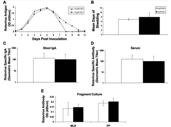
FoxP3+?regulatory T cells are not required for rotavirus clearance or rotavirus specific antibody production. FoxP3+?regulatory T cells were depleted from FoxP3DTR?mice by intraperitoneal injection of diphtheria toxin (50μg/kg of body weight). Both depleted (black bar or dashed line) and non-depleted (white bar or solid line) mice were orally inoculated with gut homogenate containing 103?ID50?of ECwt?rotavirus. Antibodies diluted in buffer containing 5% normal guinea pig serum (p/n D105-00-0050). (A) Fecal pellets were collected every day and analyzed for the presence of rotavirus by ELISA. (B) Mean days of shedding were also calculated. (C) Rotavirus specific IgA was measured in stool samples 10 dpi using ELISA. (D) Rotavirus specific antibody was measured in serum samples collected 11 dpi. (E) MLN and PP were collected 4 days post inoculation and then cultured in vitro for 4 days. Supernatants were collected and analyzed for rotavirus specific antibody using ELISA.Figure 2. PMID: 24095866.
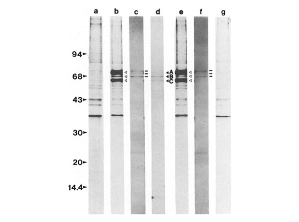
SDS-PAGE and fluorographic analysis of isoprenylated lamin polypeptides from CHO after immunoprecipitation with anti-lamin antisera. Whole cell extracts of CHO-K1 (met-18b-2) cells, labeled with 3 μCi/ml [35 S] methionine or 10 μCi/ml R-[2-14]mevalonate, were precleared by incubation with nonimmune guinea pig or human sera followed by the addition of protein A-Sepharose beads and centrifugation. The resulting supernatants were incubated with either guinea pig anti-lamin (A+B+C) antiserum or human anti-lamin (A+C) autoimmune serum. Antigen-antibody complexes were then bound to protein A-Sepharose beads, washed, eluted, and examined by SDS-PAGE and fiuorography. Fluorographic exposure was for 30 (14C-isoprenylated polypeptides) and 3 d (for [35S]methionine-labeled polypeptides). [35S]Methionine-labeled (lane b, open triangles) and 14C-isoprenylated polypeptides (lane c, bars) immunoprecipitated with guinea pig anti-lamin (A+B+C) serum. [35S]Methionine-labeled (lane e, open triangles) and 14C-isoprenylated (lane f, bars) polypeptides immunoprecipitated with human anti-lamin (A+C) autoimmune serum. [35S]Methionine-labeled polypeptides nonspecifically bound to preimmune guinea pig (lane a) and human (lane g) serum. Shown in lane d are Coomassie Blue-stained rat liver lamins A, B, and C (solid triangles). Molecular mass standards are presented to the left of the figure. Figure 5. PMID: 3170631.
|
|
|
|
Guinea Pig Serum is used as a supplement to cell culture media. Guinea Pig Serum provides a broad spectrum of macromolecules, carrier proteins for lipoid substances and trace elements, attachment and spreading factors, low molecular weight nutrients, and hormones and growth factors that promote cell growth and health. Be certain to maintain Good Cell Culture Practice, and maintain sterility of cultures that require media supplementation.
|
|
| 別品名 |
Guinea Pig serum for cell culture, cell culture grade guinea pig serum, sterile serum from guinea pig.
|
| 別包装 |
あり
|
| 種由来 |
Guinea Pig
|
| 標識物 |
Unlabeled
|
| 参考文献 |
[[Cosmo Reference Collection]][Pub Med ID]64311,65426,70481,102127,301093,343836,570843,698395,806312,1085645,2857173,2948664,3170631,3891606,4855923,6161185,6245139,6286757,6289316,6302478+他多数
|
|
| メーカー |
品番 |
包装 |
|
RKL
|
D105-00-0050
|
50 ML
|
※表示価格について
| 当社在庫 |
なし
|
| 納期目安 |
約10日
|
| 保存温度 |
-20℃
|
|


