|
※サムネイル画像をクリックすると拡大画像が表示されます。
Normal Goat Serum (NGS)
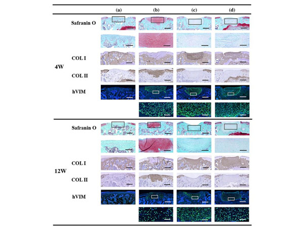
Representative microscopic images from the histological and immunohistochemical analyses. Frozen sections were blocked with 5% normal goat serum (NGS, p/n B304). Group A, untreated; Group B, LC sheet containing 5.0 × 10⁵?cells alone; Group C, SY sheet containing 5.0 × 10⁵?cells alone; and Group D, SY sheet plus LC sheet, each containing 5.0 × 10⁵?cells. For each group,?n?= 6. Four weeks after transplantation in Groups B, C, and D, the defects were filled with repaired tissue. Histological analysis of Group A revealed no Safranin O staining or formation of a chondral layer but revealed bone‐like tissue or fibrous tissue. In Group B, strong Safranin O staining was observed. In Groups C and D, no Safranin O staining was observed. Immunohistochemical analysis revealed negative staining for COL II and hVIM in Group A, positive staining for COL II and hVIM in Group B, and positive staining for COL I and hVIM in Groups C and D. At 12 weeks after transplantation, the defects in Groups B, C, and D, in which transplantation was performed, were filled with repaired tissue. Histological analysis of Group A revealed no Safranin O staining or formation of a chondral layer but revealed bone‐like tissue. In Group B, strong Safranin O staining was observed. In Groups C and D, no Safranin O staining was observed. Immunohistochemical analysis revealed negative staining for COL II and hVIM in Group A, positive staining for COL II and hVIM and partial staining for COL I in Group B, and positive staining for COL I and hVIM in Groups C and D. Low‐power images of Safranin O, COL I, COL II, and hVIM staining are shown in the upper rows (scale bar = 500 μm). High‐power images of Safranin O and hVIM are shown in the lower rows (scale bar of Safranin O = 200 μm, scale bar of hVIM = 50 μm). LC: layered chondrocyte; SY: synoviocyte; COL I: type I collagen; COL II: type II collagen; hVIM: human vimentin. Fig 3. PMID: 32652894.
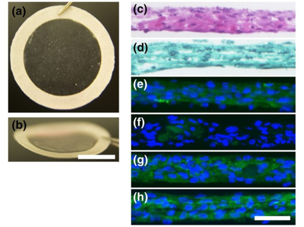
Representative macrographs and micrographs of TKA sheets. (a) Macrograph of a TKA sheet attached to a PVDF support membrane and (b) the same thin sheet seen from an angle. Scale bar?=?1?cm. Histological analysis of sections of layered chondrocyte sheets stained with (c) HE and (d) Safranin O. Immunohistochemical analysis revealed (e) positive staining for COL1, (f) slight staining for COL2, (g) positive staining for ACAN, and (h) positive staining for FN. Scale bar?=?50 μm. TKA sheets were fixed in 4% paraformaldehyde in phosphate buffer and embedded in optimal cutting temperature compound. Twenty‐micrometre‐thick sections were stained with haematoxylin and eosin (HE) or with Safranin O, Fast Green, and HE. For immunohistochemical analysis, 10‐μm sections were blocked with 5% normal goat serum (NGS; p/n B304) and 0.3% Triton X‐100 in phosphate buffer for 30?min. The sections were then incubated with primary antibodies (COL1; dilution 1:200); (COL2; dilution 1:200); (ACAN; dilution 1:10); or (FN; dilution 1:500) at 4°C overnight.? ACAN: aggrecan; COL1: Type I collagen; COL2: Type II collagen; FN: fibronectin; HE: haematoxylin and eosin; TKA: total knee arthroplasty; PVDF: polyvinylidene difluoride. Figure 1. PMID: 30058138.
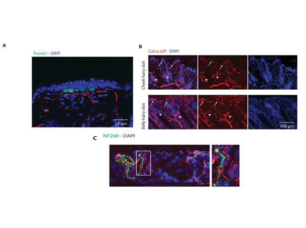
Calca-tdT neurons produce circumferential endings around hair shafts and free nerve endings in hairy skin independent of merkel cells. Hair from the skin of the back of mice was removed using a depilatory cream, and cut into square pieces of approximately 5 mm × 5 mm. The tissues were fixed in 4% PFA in PBS at 4 °C for 3?5 days. After a PBS wash, tissues were mounted in OCT medium, and sectioned at 80?90 μm on a cryostat. Skin sections were rinsed in PBS and incubated in blocking buffer (5% goat serum [p/n B304]; 0.5% Triton-X100) for 3 hours at room temperature. Sections were incubated in primary antibodies in blocking buffer at 4°C overnight. Sections were rinsed and incubated overnight in AlexaFluor conjugated secondary antibodies. (A) Immunostaining for Troma-1 (green) shows Calca labeling is independent of merkel cells. (B) Images of hairy skin from the cheek (top) and belly (bottom) from Calca-tdT mice. Calca circumferential endings (arrow heads) wrap around hair shafts while free nerve endings (arrows) are found in more superficial layers. (C) Calca-tdT free nerve endings are NF200-negative. Figure S6. PMID: 28817806.
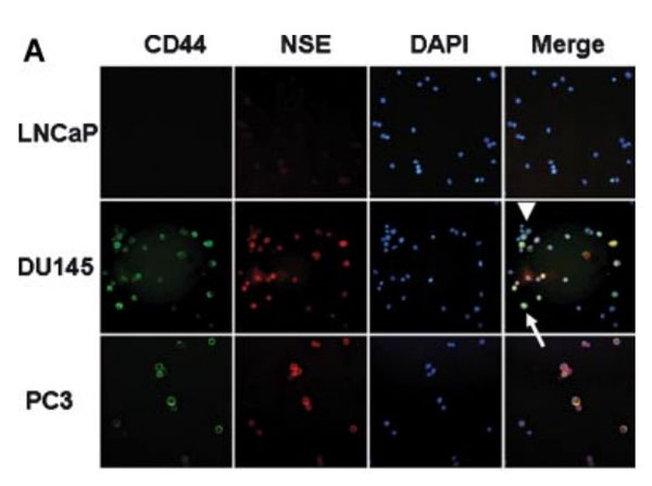
Co-expression of NSE and CD44in human prostate cancer cell lines. A:Immunofluorescence studies on cytospin samples with antibodies against CD44,NSE (with DAPI staining nuclei) show co-expression of CD44 and NSE in the same cells. LNCaP cells are double negative for the two markers and PC3 cells are double positive.ThemajorityofDU145cellsaredoublepositive (arrow) but a minority are double negative (arrowhead) (magnification 400).Cytospin preparations of PC cells were fixed in methanol for 10 min at 20C, rehydrated in PBS, and blocked in 5% normal goat serum (p/n B304) for 30 min. The slides were incubated with antibodies against CD44 (at 1:200) and neuron-specific enolase (NSE at 1:50) overnight at 4C followed by incubation with secondary antibodies (goat anti-rat IgG FITC and Alexa Fluor 546 goat anti-mouse) for 40 min at room temperature. Fig 3. PMID: 19189306.
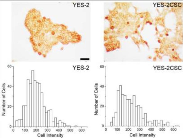
CD44 immunocytochemistry. Above:?CD44-stained cells are visible in YES-2 and in the YES-2CSC line which has high-Aldefluor staining as selected by FACS.?The cells were fixed with methanol for 5 min, washed with PBS three times, exposed to 0.3% hydrogen peroxide for 3 min, and rinsed three times with PBS. The cells were then incubated with normal goat serum (NGS, p/n B304) at 1:100 dilution for 30 min at room temperature followed by mouse monoclonal anti-human CD44 antibody at 1:10 dilution for 2 hrs at room temperature. After three rinses with PBS, the cells were incubated with goat anti-mouse horseradish peroxidase-conjugated antibody at 1:40 dilution for 30 min and then exposed to ImmunPACT diaminobenzidine for 10 min.?Below:?High CD44-staining cells are more abundant in the YES-2CSC line than in the YES-2 line. Scale bar = 100 μm.Fig 3. PMID: 23983818.
|
|
|
|
Normal Goat Serum (NGS)
|
|
| 別品名 |
blocking goat serum, blocking grade goat serum, 10% NGS, normal goat serum, goat serum blocking buffer
|
| 標識物 |
Unlabeled
|
| 精製度 |
Precipitation
|
| 発現系 |
Native
|
| その他 |
[用途]ブロッキング用
|
| 参考文献 |
[Pub Med ID]32511274
|
| [注意事項] |
濃度はロットによって異なる可能性があります。メーカーDS及びCoAからご確認ください。
|
|
| メーカー |
品番 |
包装 |
|
RKL
|
B304
|
10 ML
|
※表示価格について
| 当社在庫 |
なし
|
| 納期目安 |
2週間程度
|
| 保存温度 |
4℃
|
|
※当社では商品情報の適切な管理に努めておりますが、表示される法規制情報は最新でない可能性があります。
また法規制情報の表示が無いものは、必ずしも法規制に非該当であることを示すものではありません。
商品のお届け前に最新の製品法規制情報をお求めの際はこちらへお問い合わせください。
|
※当社取り扱いの試薬・機器製品および受託サービス・創薬支援サービス(納品物、解析データ等)は、研究用としてのみ販売しております。
人や動物の医療用・臨床診断用・食品用としては、使用しないように、十分ご注意ください。
法規制欄に体外診断用医薬品と記載のものは除きます。
|
|
※リンク先での文献等のダウンロードに際しましては、掲載元の規約遵守をお願いします。
|
|
※CAS Registry Numbers have not been verified by CAS and may be inaccurate.
|
