|
※サムネイル画像をクリックすると拡大画像が表示されます。
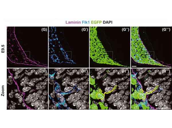
Penetration and distribution of neural crest-derived cells (NCDCs) in the?P0-Cre/EGFP?mouse telencephalon. (G?H′′′) A coronal section at the telencephalic level of the E9.5 embryo is stained with anti-laminin, Flk1, GFP antibodies and counter-stained 4′, 6-diamidino-2-phenylindole (DAPI). High-magnification images within the squares in G?G′′′ are shown in H?H′′′. Through the laminin+?basement membrane (H), an endothelial cell (white dashed line in H′) invades the telencephalon together with an EGFP+?cell (yellow line in H′′). Scale bars: (for G?G′′′), 50?μm; H′′′ (for H?H′′′), 25?μm. Figure 2. PMID: 23157329.
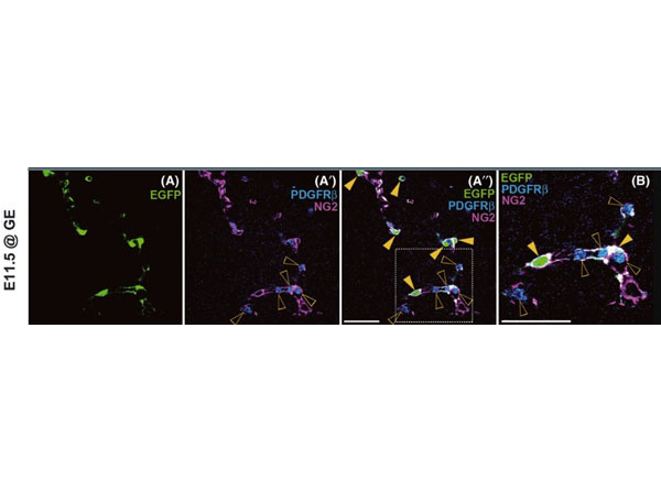
Existence of non-pericyte enhanced green fluorescent protein (EGFP+) cells in the telencephalon of E11.5?P0-Cre/EGFP?mice. (A-B) A coronal section at the telencephalic level is stained with anti-GFP, PDGFRβ, and NG2 antibodies. Most of EGFP+?cells express both PDGFRβ and NG2 (yellow arrowheads). (B) High-magnification of the dashed square in A′′. PDGFRβ is also expressed in hematopoietic cells (open arrowheads in A′?B). Fig 5. PMID: 23157329.
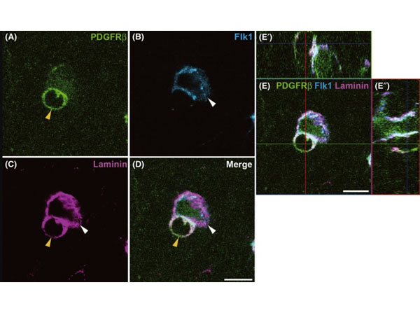
The usability of PDGFRβ and NG2 as pericyte markers in the telencephalon. (A?E′′) Immunostaining with anti-PDGFRβ, Flk1, and laminin antibodies on coronal section at the telencephalic level of E11.5?P0-Cre?mouse embryo. (A?D) A PDGFRβ+?pericyte (yellow arrowheads) wraps around Flk1+?endothelial tube (white arrowheads), and these cell types are surrounded by laminin+?basement membrane. This pattern is also clearly observed in orthogonal view of the image shown in D (E?E′′). Fig 3. PMID: 23157329.
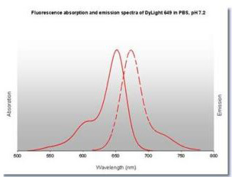
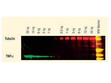
DyLight? dyes can be used for two-color Western Blot detection with low background and high signal.? Anti-tubulin was detected using a DyLight? 549 conjugate.? Anti-TNFa was detected using a DyLight? 649 conjugate. The image was captured using the Typhoon? 9410 Imaging System.
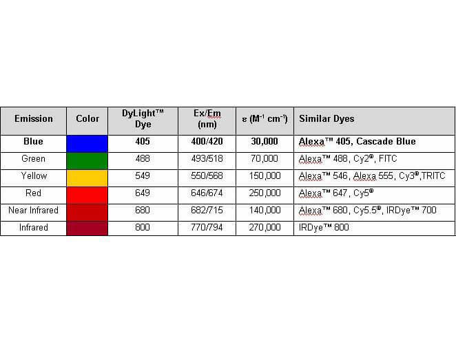
Properties of DyLight? Conjugates.
|
|
|
|
Penetration and distribution of neural crest-derived cells (NCDCs) in the?P0-Cre/EGFP?mouse telencephalon. (G?H′′′) A coronal section at the telencephalic level of the E9.5 embryo is stained with anti-laminin, Flk1, GFP antibodies and counter-stained 4′, 6-diamidino-2-phenylindole (DAPI). High-magnification images within the squares in G?G′′′ are shown in H?H′′′. Through the laminin+?basement membrane (H), an endothelial cell (white dashed line in H′) invades the telencephalon together with an EGFP+?cell (yellow line in H′′). Scale bars: (for G?G′′′), 50?μm; H′′′ (for H?H′′′), 25?μm. Figure 2. PMID: 23157329.
|
|
| 別品名 |
Goat Anti-Rat IgG DyLight 649TM Conjugated Antibody, Goat Anti-Rat IgG Antibody DyLight 649TM Conjugation
|
| 交差種 |
Rat
|
| 非交差(吸収処理)種 |
Human
Mouse
Bovine
Rabbit
Chicken
Sheep
Goat
Guinea Pig
Hamster
Equine
|
| 免疫動物 |
Goat
|
| 標識物 |
DyLightTM 649
|
| 精製度 |
Affinity Purified
|
| 参考文献 |
[Pub Med ID]23157329
|
| [注意事項] |
濃度はロットによって異なる可能性があります。メーカーDS及びCoAからご確認ください。
|
|
| メーカー |
品番 |
包装 |
|
RKL
|
612-143-120
|
100 UG
|
※表示価格について
| 当社在庫 |
なし
|
| 納期目安 |
約10日
|
| 法規制 |
毒
|
| 保存温度 |
4℃
|
|
※当社では商品情報の適切な管理に努めておりますが、表示される法規制情報は最新でない可能性があります。
また法規制情報の表示が無いものは、必ずしも法規制に非該当であることを示すものではありません。
商品のお届け前に最新の製品法規制情報をお求めの際はこちらへお問い合わせください。
|
※当社取り扱いの試薬・機器製品および受託サービス・創薬支援サービス(納品物、解析データ等)は、研究用としてのみ販売しております。
人や動物の医療用・臨床診断用・食品用としては、使用しないように、十分ご注意ください。
法規制欄に体外診断用医薬品と記載のものは除きます。
|
|
※リンク先での文献等のダウンロードに際しましては、掲載元の規約遵守をお願いします。
|
|
※CAS Registry Numbers have not been verified by CAS and may be inaccurate.
|