|
※サムネイル画像をクリックすると拡大画像が表示されます。
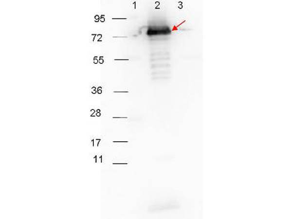
HRP-conjugated Goat-Anti-Rabbit (p/n 611-103-122) secondary antibody was used at 1:40,000 in MB-070 blocking buffer to detect a rabbit primary antibody by Western Blot. Anti-p27 antibody (200-401-C30, 1:1000 RT 30 minutes) showed detection of 0.1 μg of recombinant p27 protein. Lane 1: Molecular weight markers. Lane 2: MBP-p27 fusion protein (arrow; expected MW: 73.3 kDa). Lane 3: MBP alone. Protein was run on a 4-20% gel, then transferred to 0.45 μm nitrocellulose and blocked with 1% BSA-TTBS (p/n MB-013, diluted to 1X) overnight at 4°C. Blot was imaged on the VersaDoc MP 4000 imaging system (Bio-Rad).
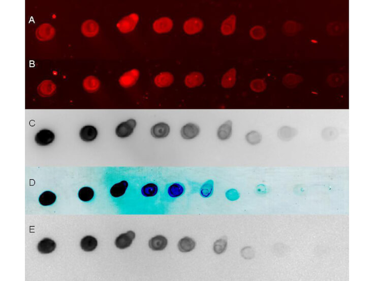
Rockland secondary antibodies detect rabbit primary antibodies in a variety of platforms. Shown here is a serial 1:1 dilution of control rabbit IgG protein (011-0102, 250ng starting total load) co incubated with Rockland HRP conjugated Goat anti Rabbit IgG (611-103-122) and Dylight 649 conjugated goat anti Rabbit (611-143-122) 1:20K in MB-070. Blot was dried and imaged (A) on Biorad Versa Doc (30 sec, DyLight649), (B) LiCor Odyssey Reader (700 nm), (C ) Rewetted incubated with Femtomax 110 reimaged using BioVersaDoc (for 60 sec), (D) Incubated with TMB substrate TMBM for 5 minutes and scanned, and (E) Rewetted for Chemiluminescence and imaged for 90 sec on the BioRad VersaDoc Imager,
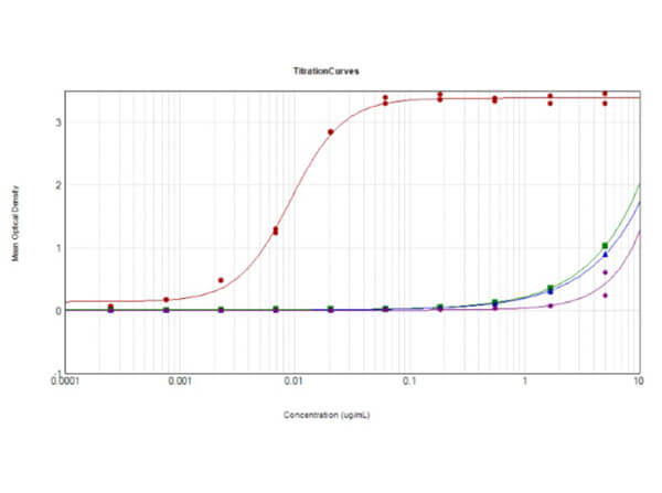
ELISA Results of Goat Anti-Rabbit IgG Antibody Peroxidase Conjugated (Min X Bv, Ch, Gt, GP, Ham, Hs, Hu, Ms, Rt, Sh Serum Proteins) tested against purified Goat Anti-Rabbit IgG Antibody HRP Conjugated MX10. Each well was coated in duplicate with 10 μg of Rabbit IgG (p/n 011-0102) [Red Line], Bovine IgG (p/n 001-0102) [Green Line], Chicken IgG (p/n 003-0102) [Blue Line], Goat IgG (p/n 005-0102) [Purple Line], Guinea Pig, Hamster, Horse, Human, Mouse, Rat, and Sheep tested with similar results. The working dilution for Rabbit IgG is 1:109,000. The starting dilution of antibody was 5μg/ml and the X-axis represents the Log10 of a 3-fold dilution. This titration is a 4-parameter curve fit where the IC50 is defined as the titer of the antibody. Assay performed using 3% Fish Gel/PBS Blocking buffer (p/n MB-066), and TMB substrate (p/n TMBE-1000).
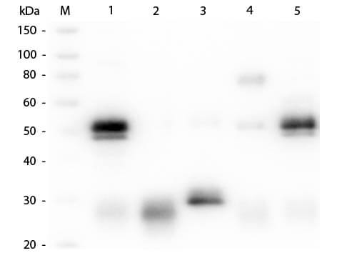
Western Blot of Unconjugated Anti-Rabbit IgG (H&L) (GOAT) Antibody (Min X Bv, Ch, Gt, GP, Ham, Hs, Hu, Ms, Rt & Sh Serum Proteins) (p/n 611-101-122). Lane M: 3 μl Molecular Ladder. Lane 1: Rabbit IgG whole molecule (p/n 011-0102). Lane 2: Rabbit IgG F(ab) Fragment (p/n 011-0105). Lane 3: Rabbit IgG F(c) Fragment (p/n 010-0103). Lane 4: Rabbit IgM Whole Molecule (p/n 011-0107). Lane 5: Normal Rabbit Serum (p/n B309). All samples were reduced. Load: 50 ng per lane. Block: MB-070 for 30 min at RT. Primary Antibody: Anti-Rabbit IgG (H&L) (GOAT) Antibody (Min X Bv, Ch, Gt, GP, Ham, Hs, Hu, Ms, Rt & Sh Serum Proteins) (p/n 611-101-122) 1:1,000 for 60 min at RT. Secondary antibody: Anti-Goat IgG (DONKEY) Peroxidase Conjugated Antibody (p/n CUST10) 1:40,000 in MB-070 for 30 min at RT. Predicted/Observed Size: 25 and 50 kDa for Rabbit IgG and Serum, 25 kDa for F(c) and F(ab), 70 and 23 kDa for IgM. Rabbit F(c) migrates slightly higher.
|
|
|
|
HRP-conjugated Goat-Anti-Rabbit (p/n 611-103-122) secondary antibody was used at 1:40,000 in MB-070 blocking buffer to detect a rabbit primary antibody by Western Blot. Anti-p27 antibody (200-401-C30, 1:1000 RT 30 minutes) showed detection of 0.1 μg of recombinant p27 protein. Lane 1: Molecular weight markers. Lane 2: MBP-p27 fusion protein (arrow; expected MW: 73.3 kDa). Lane 3: MBP alone. Protein was run on a 4-20% gel, then transferred to 0.45 μm nitrocellulose and blocked with 1% BSA-TTBS (p/n MB-013, diluted to 1X) overnight at 4°C. Blot was imaged on the VersaDoc MP 4000 imaging system (Bio-Rad).
|
|
| 別品名 |
goat anti-rabbit hrp, HRP conjugated secondary antibody, goat anti-rabbit secondary antibody, goat anti-rabbit IgG peroxidase conjugated secondary
|
| 交差種 |
Rabbit
|
| 非交差(吸収処理)種 |
Human
Mouse
Rat
Bovine
Chicken
Sheep
Goat
Guinea Pig
Hamster
Equine
|
| 適用 |
Western Blot
Enzyme Linked Immunosorbent Assay
Dot Blot
|
| 免疫動物 |
Goat
|
| 標識物 |
Horseradish Peroxidase
|
| 精製度 |
Affinity Purified
|
| 性状 |
Azide Free
|
| 参考文献 |
[Pub Med ID]32934200
|
| [注意事項] |
濃度はロットによって異なる可能性があります。メーカーDS及びCoAからご確認ください。
|
|
| メーカー |
品番 |
包装 |
|
RKL
|
611-103-122
|
1 MG
|
※表示価格について
| 当社在庫 |
なし
|
| 納期目安 |
約10日
|
| 保存温度 |
4℃
|
|
