|
※サムネイル画像をクリックすると拡大画像が表示されます。
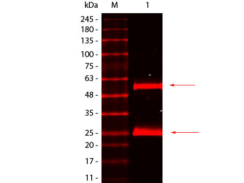
Western Blot of Goat anti-Mouse IgG Antibody DyLight 680 Conjugated Pre-absorbed. Lane 1: Mouse IgG. Load: 50 ng per lane. Primary antibody: none. Secondary antibody: Goat anti-Mouse IgG Antibody DyLight 680 Conjugated Pre-absorbed at 1:1,000 for 60 min at RT. Block: MB-070 for 30 min at RT. Predicted/Observed size: 55 kDa, 25 kDa for Mouse IgG.
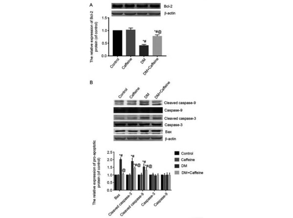
Western Blot Results using Goat Anti-Mouse IgG (H&L) Antibody DyLight? 680 Conjugated. Expression levels of Bcl-2, Bax, caspase-3 and caspase-9 proteins in the DRG. (A) The expression level of Bcl-2 protein in rat bladders was detected by western blot analysis. (B) The expression levels of Bax, caspase-3, cleaved caspase-3, caspase-9 and cleaved caspase-9 proteins in rat bladders were detected by western blot analysis.?*P<0.05 vs. control group;?#P<0.05 vs. caffeine group;?@P<0.05 vs. DM group. Bcl-2, B-cell lymphoma-2; Bax, Bcl-2-associated X protein; DRG, dorsal root ganglion; DM, diabetes mellitus. Fig 3. PMID: 33791010.
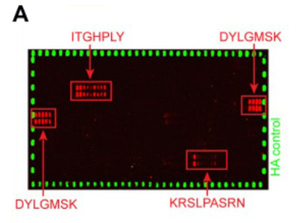
Peptide array results using Goat Anti-Mouse IgG (H&L) Antibody DyLight? 680 Conjugated. Peptide arrays identify known immunogenic epitopes in L1.(A) Synthetic 15-mer peptides with residue overlaps of 14 residues were spotted on microarrays and incubated with serum mix from five tumor-bearing animals with high titers against both L1 isoforms. Bound serum antibodies were detected with fluorophore-conjugated secondary antibodies. Fig 5. PMID: 32746966.
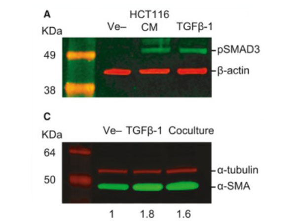
Western Blot Results using Goat Anti-Mouse IgG (H&L) Antibody DyLight? 680 Conjugated. TGF‐β‐mediated crosstalk between pericytes and CRC cells modulates pericyte secretome. (A) Incubation in HCT116 CM for 1?h induces SMAD3 phosphorylation in PC, as assessed by western blot. Exogenous recombinant TGF‐β (10?ng・mL−1) was used as a positive control, and β‐actin was used as loading control (n?=?3). (B) Confocal microscopy images of SMAD3 subcellular localization in PC cultured alone or cocultured with HCT116 cells for 48?h (n?=?3). SMAD3 is detected in the cytoplasm of PC in monoculture (arrows show nonstained nuclei). Nuclear translocation of SMAD3 takes place after coculture with HCT116 cells for 48?h (arrowheads indicate stained nuclei). HCT116 cells treated with 10?ng・mL−1?TGF‐β1 were used as a positive control. Scale bar?=?10?μm. Fig 5. PMID: 32767843.
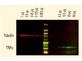
DyLight? dyes can be used for two-color Western Blot detection with low background and high signal.? Anti-tubulin was detected using a DyLight? 680 conjugate.? Anti-TNFa was detected using a DyLight? 800 conjugate. The image was captured using the OdysseyR Infrared Imaging System developed by LI-COR.
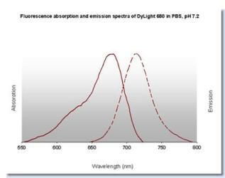
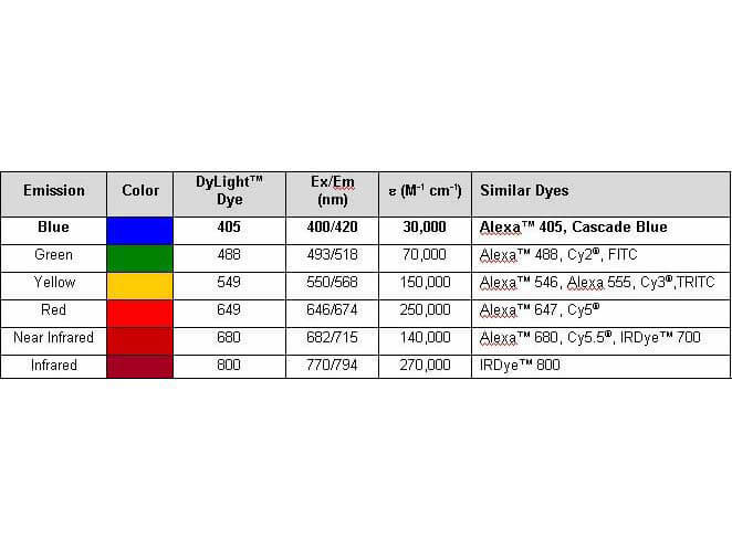
Properties of DyLight? Conjugates.
|
|
|
|
Western Blot of Goat anti-Mouse IgG Antibody DyLight 680 Conjugated Pre-absorbed. Lane 1: Mouse IgG. Load: 50 ng per lane. Primary antibody: none. Secondary antibody: Goat anti-Mouse IgG Antibody DyLight 680 Conjugated Pre-absorbed at 1:1,000 for 60 min at RT. Block: MB-070 for 30 min at RT. Predicted/Observed size: 55 kDa, 25 kDa for Mouse IgG.
|
|
| 別品名 |
Goat Anti-Mouse IgG Secondary Antibody DyLightTM680 Conjugated, Goat Anti-Mouse IgG Antibody DyLightTM680 Conjugated, Anti-mouse IgG secondary antibody, anti-mouse IgG DyLightTM680 conjugated secondary antibody
|
| 交差種 |
Mouse
|
| 非交差(吸収処理)種 |
Human
Rat
Bovine
Rabbit
Chicken
Sheep
Goat
Guinea Pig
Hamster
Equine
|
| 免疫動物 |
Goat
|
| 標識物 |
DyLightTM 680
|
| 精製度 |
Affinity Purified
|
| 参考文献 |
[Pub Med ID]28238547
|
| [注意事項] |
濃度はロットによって異なる可能性があります。メーカーDS及びCoAからご確認ください。
|
|
| メーカー |
品番 |
包装 |
|
RKL
|
610-144-121
|
100 UG
|
※表示価格について
| 当社在庫 |
なし
|
| 納期目安 |
約10日
|
| 法規制 |
毒
|
| 保存温度 |
4℃
|
|
※当社では商品情報の適切な管理に努めておりますが、表示される法規制情報は最新でない可能性があります。
また法規制情報の表示が無いものは、必ずしも法規制に非該当であることを示すものではありません。
商品のお届け前に最新の製品法規制情報をお求めの際はこちらへお問い合わせください。
|
※当社取り扱いの試薬・機器製品および受託サービス・創薬支援サービス(納品物、解析データ等)は、研究用としてのみ販売しております。
人や動物の医療用・臨床診断用・食品用としては、使用しないように、十分ご注意ください。
法規制欄に体外診断用医薬品と記載のものは除きます。
|
|
※リンク先での文献等のダウンロードに際しましては、掲載元の規約遵守をお願いします。
|
|
※CAS Registry Numbers have not been verified by CAS and may be inaccurate.
|