|
※サムネイル画像をクリックすると拡大画像が表示されます。
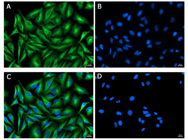
Immunofluorescence of Goat Anti-Mouse IgG (H&L) Antibody DyLight? 488 Conjugated (Min X Bv Ch Gt GP Ham Hs Hu Rb Rt & Sh Serum Proteins). Cell line:? HeLa?. Primary Antibody: Alpha Tubulin? (p/n 200-301-880?) at 4 μg/mL (1:250) for 1hr at RT?. Secondary Antibody: Goat Anti-Mouse? DyLight? 488? (p/n 610-141-121?) at 1 μg/mL (1:1000) overnight at 4 °C?. Fixative:? Ice Cold Methanol?. Permeabilization: Ice Cold Methanol?. Nuclear stain:? Hoechst 33342?. Expected Localization:? Cytoplasmic?. Image: A) Alpha Tubulin, B)? Nuclear Stain?, C) Merge?, D)? Secondary Only Control?.
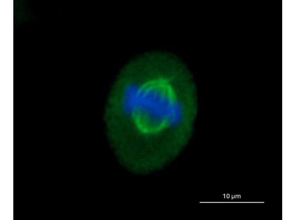
Immunofluorescence of Goat Anti-Mouse IgG (H&L) Antibody DyLight? 488 Conjugated (Min X Bv Ch Gt GP Ham Hs Hu Rb Rt & Sh Serum Proteins). Cell line:? HeLa?. Primary Antibody: Alpha Tubulin? (p/n 200-301-880?) at 4 μg/mL (1:250) for 1hr at RT?. Secondary Antibody: Goat Anti-Mouse? DyLight? 488? (p/n 610-141-121?) at 0.1μg/mL (1:10000) overnight at 4 °C?. Fixative:? Ice Cold Methanol?. Permeabilization: Ice Cold Methanol?. Nuclear stain:? Hoechst 33342?. Magnification: 40X. Expected Localization:? Cytoplasmic?. Image: HeLa cell nucleus in the anaphase stage of mitosis. Microtubule-based mitotic spindles are clearly visible.
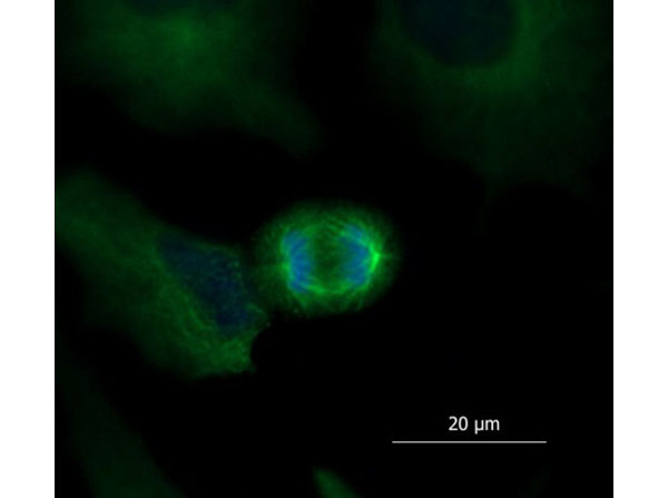
Immunofluorescence of Goat Anti-Mouse IgG (H&L) Antibody DyLight? 488 Conjugated (Min X Bv Ch Gt GP Ham Hs Hu Rb Rt & Sh Serum Proteins). Cell line:? HeLa?. Primary Antibody: Alpha Tubulin? (p/n 200-301-880?) at 4 μg/mL (1:250) for 1hr at RT?. Secondary Antibody: Goat Anti-Mouse? DyLight? 488? (p/n 610-141-121?) at 0.1 μg/mL (1:10000) overnight at 4 °C?. Fixative:? Ice Cold Methanol?. Permeabilization: Ice Cold Methanol?. Nuclear stain:? Hoechst 33342?. Magnification: 40X. Expected Localization:? Cytoplasmic?. Image: HeLa cell nucleus in the metaphase stage of mitosis. Microtubule-based mitotic spindles are clearly visible.
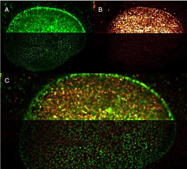
Rockland DyLight and ATTO dye conjugated antibodies provide high signal and low background for confocal microscopy and high resolution Stimulated Emission Depletion (STED) Microscopy. Both Dylight and Atto conjugated secondary antibodies maintained robust, intense signal during repeated laser excitation and de-excitation used during STED microscopy. Shown here are: A. (Green) Mouse anti NuP (NuP=Nuclear Pore Protein) detected with Dylight 488 Goat anti mouse (610-141-121) B. (Red) Rabbit Anti Ezh1/2 Pab (Ezh=enhancer of zeste homology) with detection by Rockland ATTO 425 conjugated Goat anti Rabbit (611-151-122) C. (Red and Green) Images combined. Data was collected on a STED-CW TCS-SP5 Confocal system (Leica Microsystems) equipped with a DFC 350FX Camera allowing sequential acquisition in wide-field, confocal and STED CW imaging modes and provided courtesy of: Myriam Gastard, PhD, personal communication, Leica Microsystems, Inc. USA
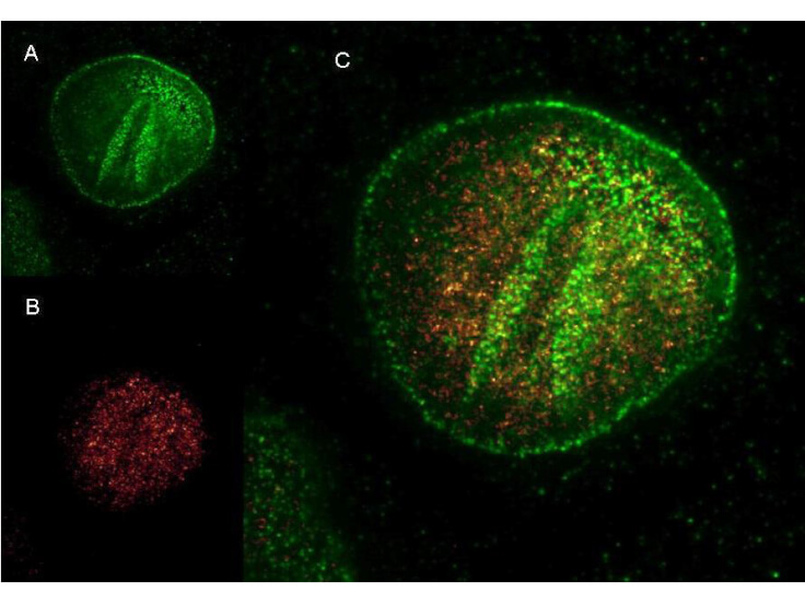
Rockland DyLight and ATTO dye conjugated antibodies provide high signal and low background for confocal microscopy (upper images) and high resolution Stimulated Emission Depletion (STED) Microscopy (lower images). Both Dylight and ATTO conjugated secondary antibodies maintained robust, intense signal during repeated laser excitation and de-excitation used during STED microscopy. Shown here are: A. (Green) Mouse anti NuP (NuP=Nuclear Pore Protein) detected with Dylight 488 Goat anti mouse (610-141-121) B. (Red) Rabbit Anti Ezh1/2 Pab (Ezh=enhancer of zeste homology) with detection by Rockland ATTO 425 conjugated Goat anti Rabbit (611-151-122) (Red and Green) Images combined. Data was collected on a STED-CW TCS-SP5 Confocal system (Leica Microsystems) equipped with a DFC 350FX Camera allowing sequential acquisition in widefield, confocal and STED CW imaging modes and provided courtesy of: Myriam Gastard, PhD, personal communication, Leica Microsystems, Inc. USA
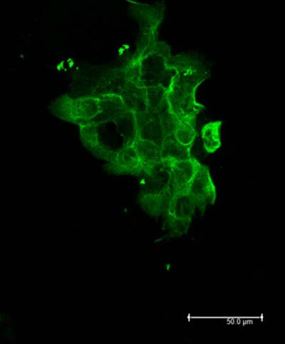
Rockland Dylight 488 Goat Anti Mouse IgG antibody-Immunofluorescence Cell Type: A431 cells Fixation: 4% paraformaldehyde 10 min Permeablization: 0.5% Triton X 30 min Primary Ab: 200-301-880 lot 28977 1:250 72 hours 4°C Secondary Ab: 610-141-121 lot 21286 1:1000 overnight 4°C
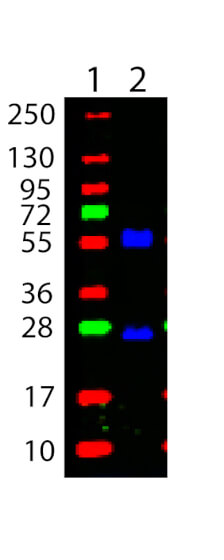
Western Blot showing detection of Mouse IgG, heavy and light chain. 100 ng of Mouse IgG (Lane 2) was run on a 4-20% gel and transferred to 0.45 μm nitrocellulose. After blocking with 1% BSA-TTBS (p/n MB-013, diluted to 1X) 30 min at 20°C, Anti-MOUSE IgG (H&L) (GOAT) Antibody DyLight? 488 Conjugated (Min X Bv Ch Gt GP Ham Hs Hu Rb Rt & Sh Serum Proteins) (p/n 610-141-121) secondary antibody was used at 1:1000 in Blocking Buffer for Fluorescent Western Blotting (p/n MB-070) and imaged using the Bio-Rad VersaDocR 4000 MP. Molecular weight markers are in lane 1.
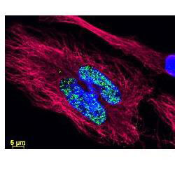
DyLight? dyes can be used for multi-color immunofluorescence microscopy with uniform fluorescence intensity throughout the image.? DyLight? dyes are exceptionally bright and photostable and are optimized for microscopy and microarray detection methods.? This image shows anti-histone detection using a DyLight? 488 conjugate (green).? Anti-Tubulin was detected using a DyLight? 549 conjugate (red). ?Nuclei were counter-stained using DAPI (blue).? The image was captured using an Axio Imager.Z1 (Zeiss Micro Imaging Inc).
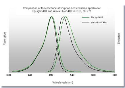
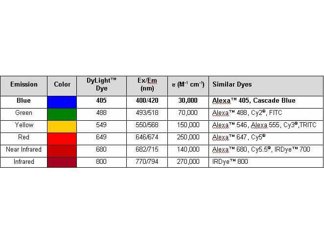
Properties of DyLight? Fluorescent Dyes.
|
|
|
|
Immunofluorescence of Goat Anti-Mouse IgG (H&L) Antibody DyLight? 488 Conjugated (Min X Bv Ch Gt GP Ham Hs Hu Rb Rt & Sh Serum Proteins). Cell line:? HeLa?. Primary Antibody: Alpha Tubulin? (p/n 200-301-880?) at 4 μg/mL (1:250) for 1hr at RT?. Secondary Antibody: Goat Anti-Mouse? DyLight? 488? (p/n 610-141-121?) at 1 μg/mL (1:1000) overnight at 4 °C?. Fixative:? Ice Cold Methanol?. Permeabilization: Ice Cold Methanol?. Nuclear stain:? Hoechst 33342?. Expected Localization:? Cytoplasmic?. Image: A) Alpha Tubulin, B)? Nuclear Stain?, C) Merge?, D)? Secondary Only Control?.
|
|
| 別品名 |
Goat Anti-Mouse IgG Secondary Antibody DyLightTM488 Conjugated, Goat Anti-Mouse IgG Antibody DyLightTM488 Conjugated, Anti-mouse IgG secondary antibody, anti-mouse IgG DyLightTM488 conjugated secondary antibody
|
| 交差種 |
Mouse
|
| 非交差(吸収処理)種 |
Human
Rat
Bovine
Rabbit
Chicken
Sheep
Goat
Guinea Pig
Hamster
Equine
|
| 適用 |
Western Blot
Dot Blot
|
| 免疫動物 |
Goat
|
| 標識物 |
DyLightTM 488
|
| 精製度 |
Affinity Purified
|
| 参考文献 |
[Pub Med ID]22675541
|
| [注意事項] |
濃度はロットによって異なる可能性があります。メーカーDS及びCoAからご確認ください。
|
|
| メーカー |
品番 |
包装 |
|
RKL
|
610-141-121
|
100 UG
|
※表示価格について
| 当社在庫 |
なし
|
| 納期目安 |
約10日
|
| 法規制 |
毒
|
| 保存温度 |
4℃
|
|
※当社では商品情報の適切な管理に努めておりますが、表示される法規制情報は最新でない可能性があります。
また法規制情報の表示が無いものは、必ずしも法規制に非該当であることを示すものではありません。
商品のお届け前に最新の製品法規制情報をお求めの際はこちらへお問い合わせください。
|
※当社取り扱いの試薬・機器製品および受託サービス・創薬支援サービス(納品物、解析データ等)は、研究用としてのみ販売しております。
人や動物の医療用・臨床診断用・食品用としては、使用しないように、十分ご注意ください。
法規制欄に体外診断用医薬品と記載のものは除きます。
|
|
※リンク先での文献等のダウンロードに際しましては、掲載元の規約遵守をお願いします。
|
|
※CAS Registry Numbers have not been verified by CAS and may be inaccurate.
|
