|
※サムネイル画像をクリックすると拡大画像が表示されます。
Western Blot analysis of IgG and IgM antibodies against?R. helvetica?whole cell antigen demonstrates the lipopolysaccaride (LPS) ladders and specific reactions against?R. helvetica?proteins in the 110?150-kDa region in serum for IgG for patients 1, 32 and 33 and for IgM for patients 3, 8, 10, 17, 20 and 22 in dilution 1:200. Lane P(h) demonstrates specific proteins and the LPS ladders reacting with a positive human serum and P(r) with a polyclonal rabbit antiserum. N(h) represent a negative human serum control. Secondary antibody Anti-human IgG DyLight?549 (p/n 609?142-123 ) and Anti-human IgM DyLight? 549 (p/n 609?142-007). Figure 1. PMID: 34712390.

Western Blot analysis of IgG antibodies against?R.?helvetica?whole cell antigen. Lane A-P demonstrates the lipopolysaccaride ladders and specific reactions against?R.?helvetica?proteins in the 110-150-kDa region for serum 2 for patients (Lane) V16(A), V43(B), V46 (C)(Area V); S71(D), S72(E), S75(F), V6(G) (Area O); K7(H), K9(I), K14(J), K46(K) K56(L) (Area K); A13(M), A16(N), A23(O), A35(P) (Area A) in titres 1:200. Lane P(h) demonstrates specific proteins and the lipopolysaccharide (LPS) ladders reacting with a human antiserum from a patient diagnosed with rickettsial infection and N(h) a healthy negative blood donor. Mw = molecular weight marker. “Fig 2” is compiled of four figure panels representing the groups of lanes that originated from different gels/blots (Gel A-D). The short vertical lines of “Fig 2” divide the individual non-adjacent lanes in the gels. The original analyses are presented in?S1?S4?Figs with Gels A-D as Supporting Information. Fig 2. PMID: 27846275.

702 Peptides are printed in duplicates randomly distributed on the microarray. Control peptides (HA and FLAG controls) are located in a square surrounding the peptides of interest. As secondary antibody DyLight? 549 conjugated goat anti-human IgG antibody and for the FLAG control peptide a mouse anti-FLAG-Cy3 antibody were used; microarrays were read using a Fujifilm Life Science FLA-5100 imaging system with a SHG 532nm (green) diode laser and an LPG filter. Fig e1. PMID: 26894206.

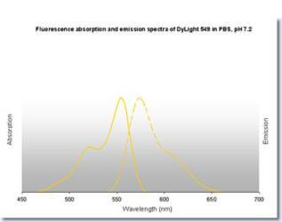
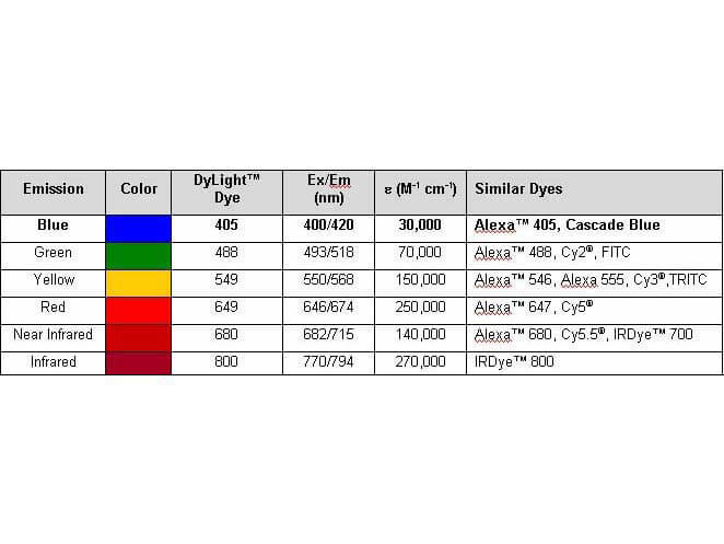
Properties of DyLight? Conjugates.
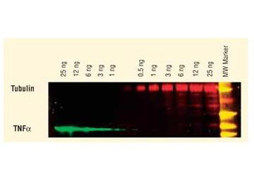
DyLight? dyes can be used for two-color western blot detection with low background and high signal. Anti-tubulin was detected using a DyLight? 549 conjugate. Anti-TNFa was detected using a DyLight? 649 conjugate. The image was captured using the Typhoon? 9410 Imaging System.
|
|
|
|
Western Blot analysis of IgG and IgM antibodies against?R. helvetica?whole cell antigen demonstrates the lipopolysaccaride (LPS) ladders and specific reactions against?R. helvetica?proteins in the 110?150-kDa region in serum for IgG for patients 1, 32 and 33 and for IgM for patients 3, 8, 10, 17, 20 and 22 in dilution 1:200. Lane P(h) demonstrates specific proteins and the LPS ladders reacting with a positive human serum and P(r) with a polyclonal rabbit antiserum. N(h) represent a negative human serum control. Secondary antibody Anti-human IgG DyLight?549 (p/n 609?142-123 ) and Anti-human IgM DyLight? 549 (p/n 609?142-007). Figure 1. PMID: 34712390.
|
|
| 別品名 |
Goat Anti Human IgG DyLight 549TM Conjugated Antibody, Goat Anti-Human IgG Antibody DyLight 549TM conjugation
|
| 交差種 |
Human
|
| 非交差(吸収処理)種 |
Mouse
Rat
Bovine
Rabbit
Chicken
Sheep
Goat
Guinea Pig
Hamster
Equine
|
| 適用 |
Enzyme Linked Immunosorbent Assay
Dot Blot
|
| 免疫動物 |
Goat
|
| 標識物 |
DyLightTM 549
|
| 精製度 |
Affinity Purified
|
| 参考文献 |
[Pub Med ID]27846275
|
| [注意事項] |
濃度はロットによって異なる可能性があります。メーカーDS及びCoAからご確認ください。
|
|
| メーカー |
品番 |
包装 |
|
RKL
|
609-142-123
|
100 UG
|
※表示価格について
| 当社在庫 |
なし
|
| 納期目安 |
約10日
|
| 法規制 |
毒
|
| 保存温度 |
4℃
|
|
※当社では商品情報の適切な管理に努めておりますが、表示される法規制情報は最新でない可能性があります。
また法規制情報の表示が無いものは、必ずしも法規制に非該当であることを示すものではありません。
商品のお届け前に最新の製品法規制情報をお求めの際はこちらへお問い合わせください。
|
※当社取り扱いの試薬・機器製品および受託サービス・創薬支援サービス(納品物、解析データ等)は、研究用としてのみ販売しております。
人や動物の医療用・臨床診断用・食品用としては、使用しないように、十分ご注意ください。
法規制欄に体外診断用医薬品と記載のものは除きます。
|
|
※リンク先での文献等のダウンロードに際しましては、掲載元の規約遵守をお願いします。
|
|
※CAS Registry Numbers have not been verified by CAS and may be inaccurate.
|