|
※サムネイル画像をクリックすると拡大画像が表示されます。
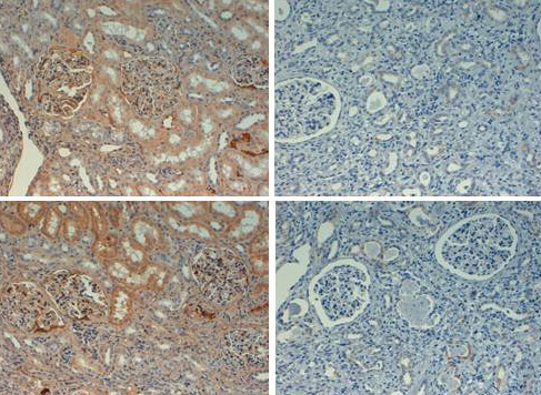
Immunohistochemistry with rabbit anti fibronectin biotin conjugated at 20X with negative controls (right). Tissue: kidney. Fixation: FFPE buffered formalin 10% conc. Antigen retrieval: Heat, Citrate pH 6.2. Pressure Cooker (top) or EDTA pH 9.5 Pressure Cooker (bottom). Primary antibody: 2ug/ml for 1 hour @ room T. Secondary antibody: Streptav. Conj. HRP 10 ug/ml circa 45 min. @ room T. Staining: antibody as precipitated red signal with a hematoxylin purple nuclear counterstain.
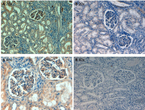
Immunohistochemistry of Rabbit Anti-Fibronectin Antibody. Tissue: human kidney at pH6 at 20x and 40x. Fixation: formalin fixed paraffin embedded. Antigen retrieval: not required. Primary antibody: Fibronectin antibody at 10 μg/mL for 1 h at RT. Secondary antibody: Peroxidase rabbit secondary antibody at 1:10,000 for 45 min at RT. Localization: Fibronectin is cytoplasmic. Staining: Fibronectin as precipitated brown signal (A) with purple nuclear counterstain. With corresponding negative control (B).
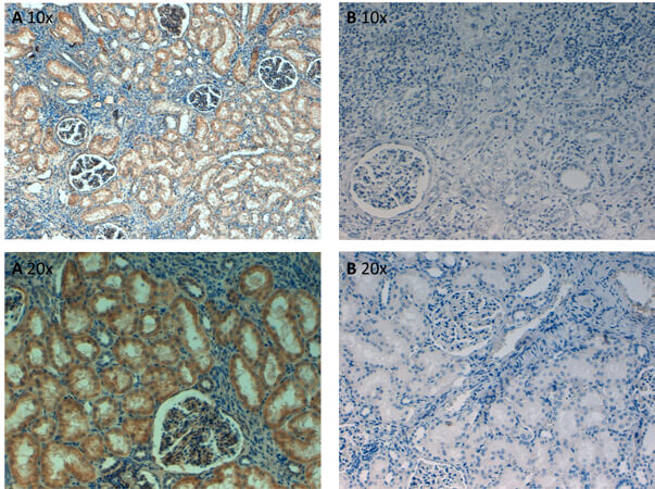
Immunohistochemistry of Rabbit Anti-Fibronectin Antibody. Tissue: human kidney at pH9 at 20x and 40x. Fixation: formalin fixed paraffin embedded. Antigen retrieval: not required. Primary antibody: Fibronectin antibody at 10 μg/mL for 1 h at RT. Secondary antibody: Peroxidase rabbit secondary antibody at 1:10,000 for 45 min at RT. Localization: Fibronectin is cytoplasmic. Staining: Fibronectin as precipitated brown signal (A) with purple nuclear counterstain. With corresponding negative control (B).
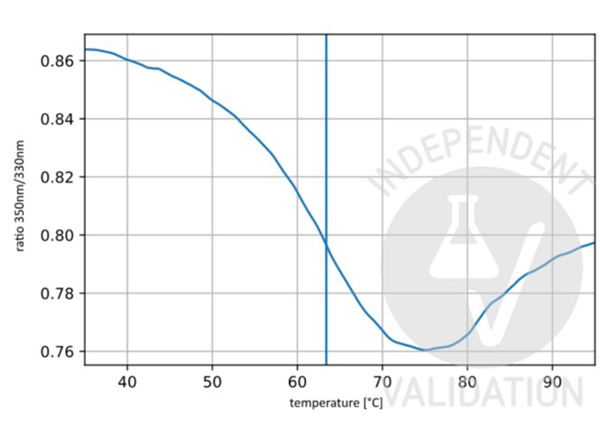
Unfolding profile of Biotin Conjugated Rabbit Anti-Fibronectin Antibody. The fluorescence signal is plotted against temperature. The vertical line indicates the Ti at 63.4°C. ?Independently Validated by?antibodies-online GmbH (p/n ABIN5596762) courtesy of NanoTemper Technologies.
|
|
|
|
Immunohistochemistry with rabbit anti fibronectin biotin conjugated at 20X with negative controls (right). Tissue: kidney. Fixation: FFPE buffered formalin 10% conc. Antigen retrieval: Heat, Citrate pH 6.2. Pressure Cooker (top) or EDTA pH 9.5 Pressure Cooker (bottom). Primary antibody: 2ug/ml for 1 hour @ room T. Secondary antibody: Streptav. Conj. HRP 10 ug/ml circa 45 min. @ room T. Staining: antibody as precipitated red signal with a hematoxylin purple nuclear counterstain.
|
|
| 別品名 |
rabbit anti-Fibronectin antibody biotin conjugation, biotin conjugated rabbit anti-Fibronectin antibody, FN1, FN, Cold-insoluble globulin, CIG, Anastellin, Ugl-Y1, Ugl-Y2, Ugl-Y3
|
| 交差種 |
Human
Mouse
Rat
Bovine
Monkey
|
| 適用 |
Western Blot
Enzyme Linked Immunosorbent Assay
Immunohistochemistry
|
| 免疫動物 |
Rabbit
|
| 標識物 |
Biotin
|
| 精製度 |
Affinity Purified
|
| GENE ID |
2335
|
| Accession No.(Gene/Protein) |
AAA53376.1, P02751
|
| Gene Symbol |
FN1
|
| 参考文献 |
Pankov and Yamada. (2002). "Fibronectin at a glance". Journal of Cell Science 115 (Pt 20): 3861-3. Cho and Mosher. (2006). "Role of fibronectin assembly in platelet thrombus formation". J. Thromb. Haemost. 4 (7): 1461-9.
|
| [注意事項] |
濃度はロットによって異なる可能性があります。メーカーDS及びCoAからご確認ください。
|
|
| メーカー |
品番 |
包装 |
|
RKL
|
600-406-117
|
0.1 MG
|
※表示価格について
| 当社在庫 |
なし
|
| 納期目安 |
約10日
|
| 法規制 |
毒
|
| 保存温度 |
4℃
|
|
※当社では商品情報の適切な管理に努めておりますが、表示される法規制情報は最新でない可能性があります。
また法規制情報の表示が無いものは、必ずしも法規制に非該当であることを示すものではありません。
商品のお届け前に最新の製品法規制情報をお求めの際はこちらへお問い合わせください。
|
※当社取り扱いの試薬・機器製品および受託サービス・創薬支援サービス(納品物、解析データ等)は、研究用としてのみ販売しております。
人や動物の医療用・臨床診断用・食品用としては、使用しないように、十分ご注意ください。
法規制欄に体外診断用医薬品と記載のものは除きます。
|
|
※リンク先での文献等のダウンロードに際しましては、掲載元の規約遵守をお願いします。
|
|
※CAS Registry Numbers have not been verified by CAS and may be inaccurate.
|
