|
※サムネイル画像をクリックすると拡大画像が表示されます。
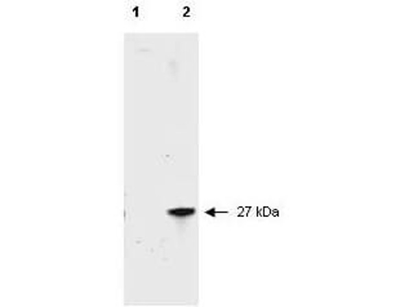
Western blot of RFP recombinant protein detected with Rockland's unconjugated polyclonal anti-RFP antibody (600-401-379). Lane 1 shows no reaction against a GFP recombinant protein present in 10 ug of HeLa cell extract. Lane 2 shows a single band detected in 10 ug of a HeLa lysate containing RFP recombinant protein. Rockland's polyclonal anti-RFP detects a 27 kDa band corresponding to the epitope tag RFP. A 4-12% Bis-Tris gradient gel (Invitrogen) was used for SDS-PAGE. The protein was transferred to nitrocellulose using standard methods. After blocking the membrane was probed with the unconjugated primary antibody diluted 1:2,500 for 1 h at room temperature followed by washes and reaction with a 1:5,000 dilution of IRDyeTM800 conjugated Goat-a-Rabbit IgG [H&L] MX (611-132-122). IRDyeTM800 fluorescence image was captured using the Odyssey(R) Infrared Imaging System developed by LI-COR. IRDye is a trademark of LI-COR, Inc. Other detection systems will yield similar results.
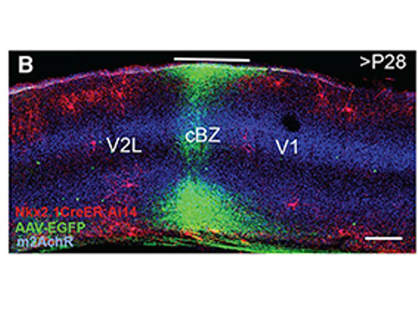
(B) Example AAV-EGFP-labeled callosal axon projection into contralateral V1/V2L border region where ChCs express RFP. The region that received callosal input is labeled as callosal binocular zone (cBZ). Fig 1. PMID:?33290732
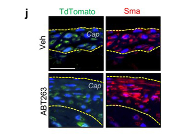
j, Representative images of tdTomato+/Sma+?cells in fibrous caps (bracketed in yellow dashed lines; quantified in?k) from indicated groups. Fig 5. PMID:?34746803
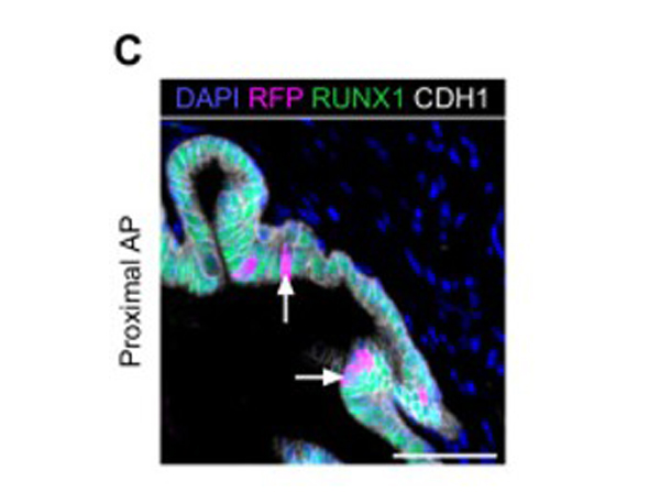
?(C) Co-immunostaining of RFP, RUNX1, CDH1 in the proximal AP. Arrows indicate RFP labeled RUNX1+?cells. Scale bar: 50 μm.? Fig 4. ?PMID:?33025905
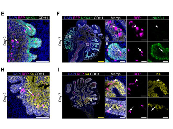
(E, F) Co-immunostaining of RFP, NKX3.1, CDH1 in UGS explants harvested at day 2 (E) and day 7 (F). Higher magnification images of (i) proximal and (ii) distal regions are shown for day 7. Arrows show RFP+?NKX3.1+?cells, arrowheads show RFP+?NKX3.1-?cells. Scale bars: 200 μm (yellow) and 50 μm (white). (H, I) Co-immunostaining of RFP, K4, CDH1 in UGS explants harvested at day 2 (H) and day 7 (I). Higher magnification images of (i) proximal and (ii) distal regions are shown for day 7. Arrows show RFP+?K4 cells, arrowheads show RFP+?K4+?cells. Scale bars: 200 μm (yellow) and 50 μm (white). Fig 7. ?PMID:?33025905
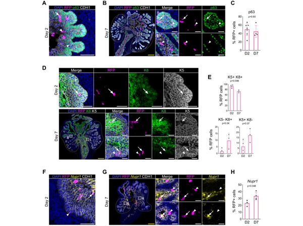
(A, B) Co-immunostaining of RFP, p63, CDH1 in UGS explants harvested at day 2 (A) and day 7 (B). Higher magnification images of (i) proximal and (ii) distal regions are shown for day 7. Arrows show RFP+?p63+?cells, arrowheads show RFP+?p63-?cells. Scale bars: 200 μm (yellow) and 50 μm (white). (C) Quantification of the percentage of epithelial RFP+?p63+?cells at day 2 (n?=?7) and day 7 (n?=?4) of UGS explant cultures. Quantification was performed within the boundaries delimited in?B?by dotted lines. (D) Co-immunostaining of RFP, K5, K8 in UGS explants harvested at day 2 (top) and day 7 (bottom). Higher magnification images of proximal (i) and (ii) distal regions are shown for day 7. Arrows show RFP+?K5+?K8+?cells, chevron arrows show RFP+?K5-?K8+, arrowheads show RFP+?K5+?K8-?cells. Scale bars: 200 μm (yellow) and 50 μm (white). (E) Quantification of the percentage of epithelial K5+?K8+, K5-?K8+?cells and K5+?K8-?cells in the RFP subset at day 2 (n?=?4) and day 7 (n?=?3) of UGS explant cultures. Quantifications were performed within the boundaries delimited in?D?by dotted lines. (F, G) Co-immunostaining of RFP,?Nupr1?(mRNA), CDH1 in UGS explants harvested at day 2 (F) and day 7 (G). Higher magnification images of (i) proximal and (ii) distal regions are shown for day 7. Arrows show RFP+?Nupr1+?cells, arrowheads show RFP+?Nupr1-?cells. Scale bars: 200 μm (yellow) and 50 μm (white). (J) Quantification of the percentage of epithelial K4+?cells in the RFP subset at day 2 (n?=?3) and day 7 (n?=?4) of UGS explant cultures. Quantification was performed within the boundaries delimited in?G?by dotted lines. Source files are available in?Figure 7―figure supplement 1―source data 1. ?PMID:?33025905
|
|
|
|
Western blot of RFP recombinant protein detected with Rockland's unconjugated polyclonal anti-RFP antibody (600-401-379). Lane 1 shows no reaction against a GFP recombinant protein present in 10 ug of HeLa cell extract. Lane 2 shows a single band detected in 10 ug of a HeLa lysate containing RFP recombinant protein. Rockland's polyclonal anti-RFP detects a 27 kDa band corresponding to the epitope tag RFP. A 4-12% Bis-Tris gradient gel (Invitrogen) was used for SDS-PAGE. The protein was transferred to nitrocellulose using standard methods. After blocking the membrane was probed with the unconjugated primary antibody diluted 1:2,500 for 1 h at room temperature followed by washes and reaction with a 1:5,000 dilution of IRDyeTM800 conjugated Goat-a-Rabbit IgG [H&L] MX (611-132-122). IRDyeTM800 fluorescence image was captured using the Odyssey(R) Infrared Imaging System developed by LI-COR. IRDye is a trademark of LI-COR, Inc. Other detection systems will yield similar results.
|
|
| 別品名 |
rabbit anti-RFP antibody fluorescein conjugation, FITC conjugated rabbit anti-RFP antibody, DsRed, rDsRed, Discosoma sp. Red Fluorescent Protein, Red fluorescent protein drFP583
|
| 非交差(吸収処理)種 |
Human
Mouse
Rat
|
| 適用 |
Dot Blot
|
| 免疫動物 |
Rabbit
|
| 標識物 |
Fluorescein Isothiocyanate
|
| 精製度 |
Affinity Purified
|
| Accession No.(Gene/Protein) |
Q9U6Y8
|
| Gene Symbol |
DsRed
|
| Tag情報 |
RFP
|
| 参考文献 |
[[Cosmo Reference Collection]][Pub Med ID]33025905,33290732,34746803,39505408
|
| [注意事項] |
濃度はロットによって異なる可能性があります。メーカーDS及びCoAからご確認ください。
|
|
| メーカー |
品番 |
包装 |
|
RKL
|
600-402-379
|
100 UG
|
※表示価格について
| 当社在庫 |
なし
|
| 納期目安 |
約10日
|
| 法規制 |
毒
|
| 保存温度 |
4℃
|
|
※当社では商品情報の適切な管理に努めておりますが、表示される法規制情報は最新でない可能性があります。
また法規制情報の表示が無いものは、必ずしも法規制に非該当であることを示すものではありません。
商品のお届け前に最新の製品法規制情報をお求めの際はこちらへお問い合わせください。
|
※当社取り扱いの試薬・機器製品および受託サービス・創薬支援サービス(納品物、解析データ等)は、研究用としてのみ販売しております。
人や動物の医療用・臨床診断用・食品用としては、使用しないように、十分ご注意ください。
法規制欄に体外診断用医薬品と記載のものは除きます。
|
|
※リンク先での文献等のダウンロードに際しましては、掲載元の規約遵守をお願いします。
|
|
※CAS Registry Numbers have not been verified by CAS and may be inaccurate.
|


