|
※サムネイル画像をクリックすると拡大画像が表示されます。
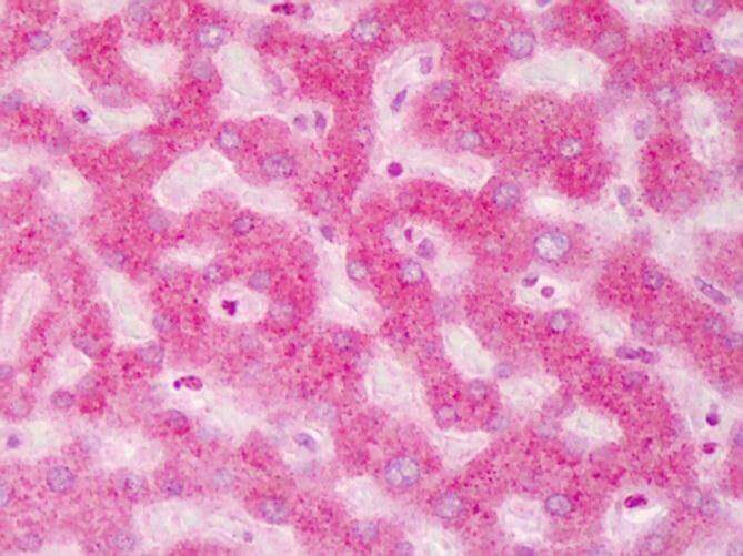
Immunohistochemistry of ABCB5 antibody. Tissue: human liver. Fixation: formalin fixed paraffin embedded. Antigen retrieval: user optimized. Primary antibody: ABCB5 at 1:200. Secondary antibody: Peroxidase goat anti-rabbit at 1:10,000 for 45 min at RT. Localization: Moderate to strong cytoplasmic and membranous staining was observed in hepatocytes. Occasional nuclear staining was observed in hepatocytes and sinusoidal cells. Staining: antibody as precipitated red signal with a hematoxylin purple nuclear counterstain.
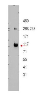
Western blot using Rockland's affinity purified anti-ABCB5 antibody shows detection of ABCB5 beta in ~12.5 ug of transfected-Hi5 whole cell lysate. No reaction was seen when antibody was pre-incubated with the immunizing peptide (data not shown). A 3-8% Tris-acetate gel was used for separation. The arrowhead corresponds to 117 kDa ABCB5. The membrane was probed with the primary antibody at a 1:10,000 dilution in 5% milk in TBST at 4° C, overnight. Personal Communication, JP Gillet, CCR-NCI, Bethesda, MD.
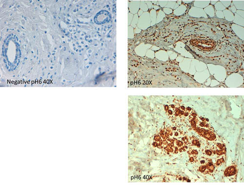
Immunohistochemistry of Rabbit anti ABCB5 antibody. Tissue: human breast carcinoma at pH6. (left, neg control) (right, 20X, 40X). Fixation: formalin fixed paraffin embedded. Primary antibody: ABCB5 antibody at 10 μg/mL for 1 h at RT. Secondary antibody: Peroxidase rabbit secondary antibody at 1:10,000 for 45 min at RT. Localization: ABCB5 is cytoplasmic. Staining: ABCB5 as precipitated brown signal with hematoxylin purple nuclear counterstain.
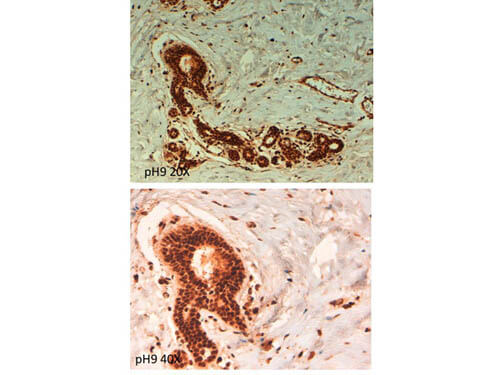
Immunohistochemistry of Rabbit anti ABCB5 antibody. Tissue: human breast carcinoma at pH9. (20X, 40X) Fixation: formalin fixed paraffin embedded. Primary antibody: ABCB5 antibody at 10 μg/mL for 1 h at RT. Secondary antibody: Peroxidase rabbit secondary antibody at 1:10,000 for 45 min at RT. Localization: ABCB5 is cytoplasmic. Staining: ABCB5 as precipitated brown signal.
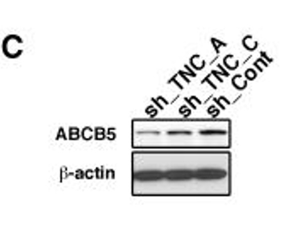
(C) Immunoblot showing that protein expression of ABCB5 is decreased in WM3734 cells transduced with TNC shRNA (sh_TNC_A and sh_TNC_C) when compared to non-targeting shRNA (sh_Cont). β-actin indicates equal loading.? Fig 5. PMID:?20729912
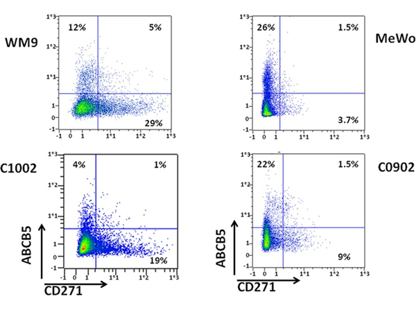
Analysis of ABCB5 and CD271 populations in melanomas. WM9, MeWo, C1002 and C0902 were labeled for CD271 and ABCB5. Cells were analyzed by flow cytometry. CD271 intensity was plotted on abscissa and ABCB5 on ordinate. Fig 1. PMID:?25105565
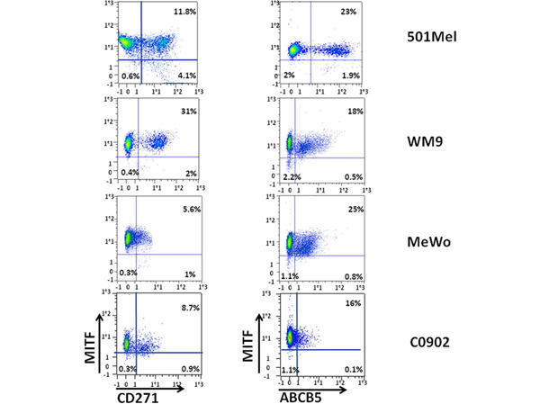
Analysis of MITF expression in CD271 and ABCB5 populations. WM9, 501-Mel, MeWo and C0902 were labeled for CD271 and MITF (left panel) or ABCB5 and MITF (right panel). CD271 or ABCB5 intensity was plotted on abscissa, and MITF expression on ordinate. Fig 2. PMID:?25105565
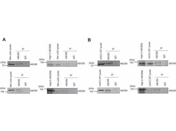
Coimmunoprecipitation of ABCB5β-ABCB6 and ABCB5β-ABCB9 demonstrates the presence of these heterodimers in Mel JuSo and UACC-257?cells.?The Mel JuSo (A) or UACC-257 (B) proteins were immunoprecipitated (IP) with either an anti-ABCB5, anti-ABCB6, or anti-ABCB9 antibody. The precipitated proteins were revealed by Western blotting after SDS-PAGE using the corresponding antibody indicated on the?right side?of each blot. Fifteen micrograms of total proteins from the starting cell lysate were loaded in the first lane, while the total IP eluate was loaded on the gel. ABCB5β was expressed in High-Five insect cells, and total membrane proteins were prepared and loaded on the gel (High5 ABCB5β) as a complementary molecular weight marker when using anti-ABCB5. An isotype control was performed to determine the specificity of the signal obtained in Western blot. Fig 3. PMID:?38145744
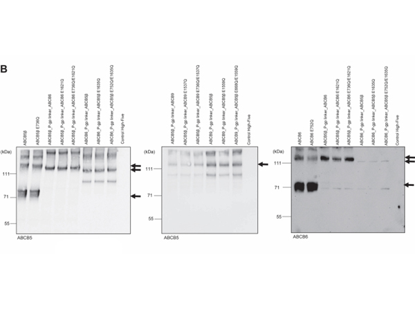
Expression of ABCB5β, ABCB6, ABCB5β_P-gp linker_ABCB6, ABCB6_P-gp linker_ABCB5β, ABCB9, ABCB5β_P-gp linker_ABCB9, and ABCB9_P-gp linker_ABCB5β in High-Five insect cells.?Total membranes vesicles prepared from High-Five cells infected with baculovirus containing either ABCB5β, ABCB6, ABCB9, ABCB5β_P-gp linker_ABCB6, ABCB6_P-gp linker_ABCB5β, ABCB5β_P-gp linker_ABCB9, ABCB9_P-gp linker_ABCB5β and their corresponding EQ mutants were subjected to SDS-PAGE, followed by Western blotting with anti-ABCB5 and anti-ABCB6 antibodies (B).?Bands of interest are highlighted by?black arrows. Fig 5. PMID:?38145744
|
|
|
|
Immunohistochemistry of ABCB5 antibody. Tissue: human liver. Fixation: formalin fixed paraffin embedded. Antigen retrieval: user optimized. Primary antibody: ABCB5 at 1:200. Secondary antibody: Peroxidase goat anti-rabbit at 1:10,000 for 45 min at RT. Localization: Moderate to strong cytoplasmic and membranous staining was observed in hepatocytes. Occasional nuclear staining was observed in hepatocytes and sinusoidal cells. Staining: antibody as precipitated red signal with a hematoxylin purple nuclear counterstain.
|
|
| 別品名 |
rabbit anti-ABCB5 Antibody, ABCB 5, ABCB-5, ATP binding cassette sub family B (MDR/TAP) member 5 antibody, ATP binding cassette sub family B member 5 antibody, EST422562 antibody, P glycoprotein ABCB5 antibody
|
| 別包装 |
あり
|
| 交差種 |
Human
|
| 適用 |
Western Blot
Enzyme Linked Immunosorbent Assay
Immunohistochemistry
|
| 免疫動物 |
Rabbit
|
| 標識物 |
Unlabeled
|
| 精製度 |
Affinity Purified
|
| GENE ID |
340273
|
| Accession No.(Gene/Protein) |
56849536, Q2M3G0
|
| Gene Symbol |
ABCB5
|
| 参考文献 |
[[Cosmo Reference Collection]][Pub Med ID]20729912,25105565,38145744
|
| [注意事項] |
濃度はロットによって異なる可能性があります。メーカーDS及びCoAからご確認ください。
|
|
| メーカー |
品番 |
包装 |
|
RKL
|
600-401-A77
|
100 UG
|
※表示価格について
| 当社在庫 |
なし
|
| 納期目安 |
約10日
|
| 保存温度 |
-20℃
|
|
※当社では商品情報の適切な管理に努めておりますが、表示される法規制情報は最新でない可能性があります。
また法規制情報の表示が無いものは、必ずしも法規制に非該当であることを示すものではありません。
商品のお届け前に最新の製品法規制情報をお求めの際はこちらへお問い合わせください。
|
※当社取り扱いの試薬・機器製品および受託サービス・創薬支援サービス(納品物、解析データ等)は、研究用としてのみ販売しております。
人や動物の医療用・臨床診断用・食品用としては、使用しないように、十分ご注意ください。
法規制欄に体外診断用医薬品と記載のものは除きます。
|
|
※リンク先での文献等のダウンロードに際しましては、掲載元の規約遵守をお願いします。
|
|
※CAS Registry Numbers have not been verified by CAS and may be inaccurate.
|


