|
※サムネイル画像をクリックすると拡大画像が表示されます。
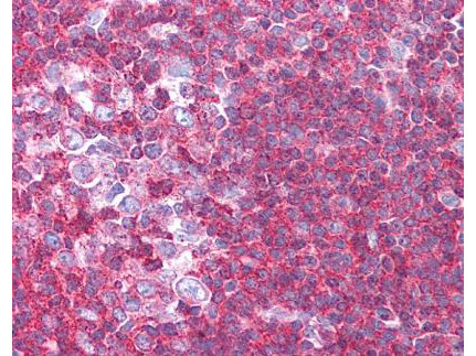
Rockland's affinity purified anti-Sipa1 antibody was used at 1.25 ug/ml to detect signal in a variety of tissues including multi-human, multi-brain and multi-cancer slides. This image shows moderate to strong positive staining of lymphocytes within human tonsil at 40X. Tissue was formalin-fixed and paraffin embedded. The image shows localization of the antibody as the precipitated red signal, with a hematoxylin purple nuclear counterstain. Personal Communication, Tina Roush, LifeSpanBiosciences, Seattle, WA.
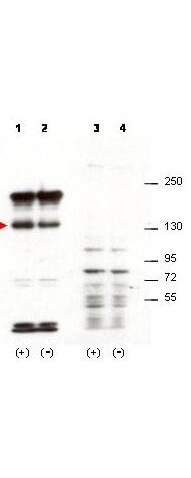
Western blot using Rockland's affinity purified anti-Sipa1 antibody shows detection of over-expressed Sipa1 in lysates from mouse 3T3 cells transfected with Sipa1 (lane 1). Endogenous Sipa1 is detected in lane 2, which contains lysate from 3T3 cells mock-transfected with LacZGLB, although at a significantly reduced level compared to transfected cells. Lane 3 and 4 are similar to lanes 1 and 2 except the antibody was preincubated with the immunizing peptide prior to reaction with the membrane. The identity of the higher and lower molecular weight bands is unknown. The band at ~130 kDa, indicated by the arrowhead, corresponds to recombinant Sipa1. Primary antibody was used at 1:1250. Personal communication, H. Yang, L. Lukes and K. Hunter, NCI, Bethesda, MD.
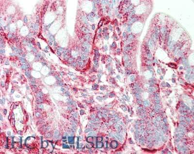
Immunohistochemistry of rabbit anti-Sipa1 antibody. Tissue: small intestine. Fixation: formalin fixed paraffin embedded. Antigen retrieval: not required. Primary antibody: Anti-Sipa1 at 5 μg/mL for 1 h at RT. Secondary antibody: Peroxidase rabbit secondary antibody at 1:10,000 for 45 min at RT. Staining: Sipa-1 as precipitated red signal with hematoxylin purple nuclear counterstain.
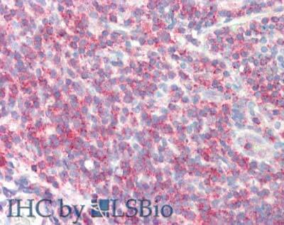
Immunohistochemistry of rabbit anti-Sipa1 antibody. Tissue: tonsil. Fixation: formalin fixed paraffin embedded. Antigen retrieval: not required. Primary antibody: Anti-Sipa1 at 5 μg/mL for 1 h at RT. Secondary antibody: Peroxidase rabbit secondary antibody at 1:10,000 for 45 min at RT. Staining: Sipa-1 as precipitated red signal with hematoxylin purple nuclear counterstain.
|
|
|
|
Rockland's affinity purified anti-Sipa1 antibody was used at 1.25 ug/ml to detect signal in a variety of tissues including multi-human, multi-brain and multi-cancer slides. This image shows moderate to strong positive staining of lymphocytes within human tonsil at 40X. Tissue was formalin-fixed and paraffin embedded. The image shows localization of the antibody as the precipitated red signal, with a hematoxylin purple nuclear counterstain. Personal Communication, Tina Roush, LifeSpanBiosciences, Seattle, WA.
|
|
| 別品名 |
rabbit anti-Sipa1 antibody, Sipa-1, Sipa 1, GTPase activating protein Spa 1 antibody, p130 SPA-1 antibody, Signal induced proliferation associated 1 antibody, SPA1
|
| 交差種 |
Human
Mouse
|
| 適用 |
Western Blot
Enzyme Linked Immunosorbent Assay
Immunohistochemistry
|
| 免疫動物 |
Rabbit
|
| 抗原部位 |
N-terminus
|
| 標識物 |
Unlabeled
|
| 精製度 |
Affinity Purified
|
| GENE ID |
20469
|
| Accession No.(Gene/Protein) |
AAH54824.1, P46062
|
| Gene Symbol |
Sipa1
|
| [注意事項] |
濃度はロットによって異なる可能性があります。メーカーDS及びCoAからご確認ください。
|
|
| メーカー |
品番 |
包装 |
|
RKL
|
600-401-A36
|
100 UG
|
※表示価格について
| 当社在庫 |
なし
|
| 納期目安 |
約10日
|
| 保存温度 |
-20℃
|
|
※当社では商品情報の適切な管理に努めておりますが、表示される法規制情報は最新でない可能性があります。
また法規制情報の表示が無いものは、必ずしも法規制に非該当であることを示すものではありません。
商品のお届け前に最新の製品法規制情報をお求めの際はこちらへお問い合わせください。
|
※当社取り扱いの試薬・機器製品および受託サービス・創薬支援サービス(納品物、解析データ等)は、研究用としてのみ販売しております。
人や動物の医療用・臨床診断用・食品用としては、使用しないように、十分ご注意ください。
法規制欄に体外診断用医薬品と記載のものは除きます。
|
|
※リンク先での文献等のダウンロードに際しましては、掲載元の規約遵守をお願いします。
|
|
※CAS Registry Numbers have not been verified by CAS and may be inaccurate.
|
