

| 別品名 |
GTPase activating protein Spa 1 antibody, MGC102688 antibody, MGC17037 antibody, p130 SPA1 antibody, Signal induced proliferation associated 1 antibody
|
| 抗原部位 |
N-terminus
|
| 種由来 |
Mouse
|
| 標識物 |
Unlabeled
|
| 精製度 |
Affinity Purified
|
| 適用 |
Western Blot
Enzyme Linked Immunosorbent Assay
Immunohistochemistry
|
| 免疫動物 |
Rabbit
|
| 交差種 |
Human
Mouse
|
| GENE ID |
20469
|
| Accession No.(Gene/Protein) |
AAH54824, P46062
|
| Gene Symbol |
SIPA1
|
| 形状 |
滅菌済み液状品
|
| [注意事項] |
濃度はロットによって異なる可能性があります。メーカーDS及びCoAからご確認ください。
|
|
※サムネイル画像をクリックすると拡大画像が表示されます。
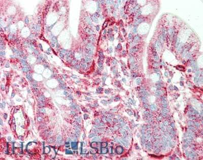
Immunohistochemistry of rabbit anti Sipa1 antibody. Tissue: small intestine. Fixation: formalin fixed paraffin embedded. Antigen retrieval: not required. Primary antibody: Anti Sipa1 at 5 ug/mL for 1 h at RT. Secondary antibody: Peroxidase rabbit secondary antibody at 1:10,000 for 45 min at RT. Staining: Sipa 1 as precipitated red signal with hematoxylin purple nuclear counterstain.
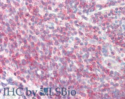
Immunohistochemistry of rabbit anti-Sipa1 antibody. Tissue: tonsil. Fixation: formalin fixed paraffin embedded. Antigen retrieval: not required. Primary antibody: Anti-Sipa1 at 5 ug/mL for 1 h at RT. Secondary antibody: Peroxidase rabbit secondary antibody at 1:10,000 for 45 min at RT. Staining: Sipa-1 as precipitated red signal with hematoxylin purple nuclear counterstain.
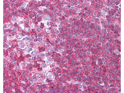
Rockland's affinity purified anti-Sipa1 antibody was used at 1.25 ug/ml to detect signal in a variety of tissues including multi-human, multi-brain and multi-cancer slides. This image shows moderate to strong positive staining of lymphocytes within human tonsil at 40X. Tissue was formalin-fixed and paraffin embedded. The image shows localization of the antibody as the precipitated red signal, with a hematoxylin purple nuclear counterstain. Personal Communication, Tina Roush, LifeSpanBiosciences, Seattle, WA.
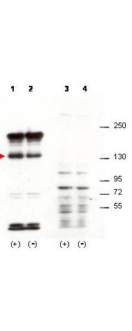
Western blot using Rockland's affinity purified anti-Sipa1 antibody shows detection of over-expressed Sipa1 in lysates from mouse 3T3 cells transfected with Sipa1 (lane 1). Endogenous Sipa1 is detected in lane 2, which contains lysate from 3T3 cells mock-transfected with LacZGLB, although at a significantly reduced level compared to transfected cells. Lane 3 and 4 are similar to lanes 1 and 2 except the antibody was preincubated with the immunizing peptide prior to reaction with the membrane. The identity of the higher and lower molecular weight bands is unknown. The band at ~130 kDa, indicated by the arrowhead, corresponds to recombinant Sipa1. Primary antibody was used at 1:1250. Personal communication, H. Yang, L. Lukes and K. Hunter, NCI, Bethesda, MD.
|
|
|
|
Immunohistochemistry of rabbit anti Sipa1 antibody. Tissue: small intestine. Fixation: formalin fixed paraffin embedded. Antigen retrieval: not required. Primary antibody: Anti Sipa1 at 5 ug/mL for 1 h at RT. Secondary antibody: Peroxidase rabbit secondary antibody at 1:10,000 for 45 min at RT. Staining: Sipa 1 as precipitated red signal with hematoxylin purple nuclear counterstain.
|
|
|
| メーカー |
品番 |
包装 |
|
RKL
|
600-401-A36
|
100 UG
|
※表示価格について
| 当社在庫 |
なし
|
| 納期目安 |
約10日
|
| 保存温度 |
-20℃
|
|

