|
※サムネイル画像をクリックすると拡大画像が表示されます。
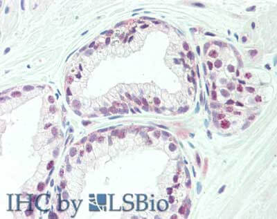
Immunohistochemistry of rabbit anti-TAF1 antibody. Tissue: prostate. Fixation: formalin fixed paraffin embedded. Antigen retrieval: not required. Primary antibody: Anti-TAF1 at 10 μg/mL for 1 h at RT. Secondary antibody: Peroxidase rabbit secondary antibody at 1:10,000 for 45 min at RT. Staining: TAF-1 as precipitated red signal with hematoxylin purple nuclear counterstain.
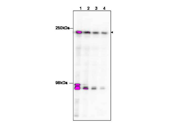
Western blot using Rockland's affinity purified anti-TAF1 to detect TAF1 in HeLa nuclear extract (arrowhead). The membrane was probed with the primary antibody at dilutions of 1:100 (lane 1), 1:250 (lane 2), 1: 500 (Lane 3 and 1:1,000 (Lane 4). The identity of the bands at ~95 kDa is unknown, but may be degraded TAF1. Personal Communication, Anne Gegonne, CCR-NCI, Bethesda, MD.
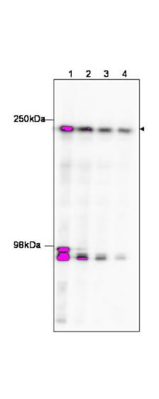
Western blot using Rockland's affinity purified anti-TAF1 to detect TAF1 in HeLa nuclear extract (arrowhead). The membrane was probed with the primary antibody at dilutions of 1:100 (lane 1), 1:250 (lane 2), 1: 500 (Lane 3 and 1:1,000 (Lane 4). The identity of the bands at ~95 kDa is unknown, but may be degraded TAF1. Personal Communication, Anne Gegonne, CCR-NCI, Bethesda, MD.
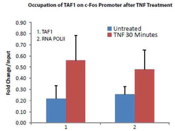
Transcription Initiation Factor TFIID Subunit 1 (TAF1) antibody was used to detect TAF1 in treated and untreated HeLa Cells. HeLa cells were treated with TNF alpha and Chromatin was prepared by EZ Magna Chip Kit (Millipore). CHIP was performed on fos promoters using 5 μg of TAF1 antibody from Rockland (P/n 600-401-995) and an RNA PolII antibody. Image with data provided courtesy of Shiraz Mujtaba, Ph.D., Dept. of Structural & Chemical Biology, Mount Sinai School of Medicine.
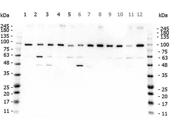
Western Blot of Rabbit anti-TAF1 antibody. Marker: Opal Pre-stained ladder (p/n MB-210-0500). Lane 1: HEK293 lysate (p/n W09-000-365). Lane 2: HeLa Lysate (p/n W09-000-363). Lane 3: MCF-7 Lysate (p/n W09-000-360). Lane 4: Jurkat Lysate (p/n W09-000-370). Lane 5: A431 Lysate (p/n W09-000-361). Lane 6: A549 Lysate (p/n W09-001-372). Lane 7: LNCap Lysate (p/n W09-001-GJ9). Lane 8: MOLT-4 Lysate (p/n W09-001-GK2). Lane 9: Ramos Lysate (p/n W09-000-GK4). Lane 10: Raji Lsyate (p/n W09-001-368). Lane 11: A-172 Lysate (p/n W09-001-GL5). Lane 12: NIH/3T3 Lysate (p/n W10-000-358). Load: 35 μg per lane. Primary antibody: TAF1 antibody at 0.2ug/mL overnight at 4C. Secondary antibody: Peroxidase rabbit secondary antibody (p/n 611-103-122) at 1:30,000 for 60 min at RT. Blocking Buffer: 1% Casein-TTBS for 30 min at RT. Predicted/Observed size: 250kDa for TAF1.
|
|
|
|
Immunohistochemistry of rabbit anti-TAF1 antibody. Tissue: prostate. Fixation: formalin fixed paraffin embedded. Antigen retrieval: not required. Primary antibody: Anti-TAF1 at 10 μg/mL for 1 h at RT. Secondary antibody: Peroxidase rabbit secondary antibody at 1:10,000 for 45 min at RT. Staining: TAF-1 as precipitated red signal with hematoxylin purple nuclear counterstain.
|
|
| 別品名 |
Rabbit anti-TAF1 antibody, rabbit anti-Anti-Transcription Initiation Factor TFIID Subunit 1 antibody, TAF-1, TAF 1, Cell cycle gene 1 protein, TBP-associated factor 250 kDa, TAFII-250
|
| 交差種 |
Human
Mouse
|
| 適用 |
Western Blot
Enzyme Linked Immunosorbent Assay
Immunohistochemistry
Chromatin Immunoprecipitation
|
| 免疫動物 |
Rabbit
|
| 抗原部位 |
C-terminus
|
| 標識物 |
Unlabeled
|
| 精製度 |
Affinity Purified
|
| GENE ID |
6872
|
| Accession No.(Gene/Protein) |
NP_001273003.1, P21675
|
| Gene Symbol |
TAF1
|
| 参考文献 |
[Pub Med ID]22411629
|
|
| メーカー |
品番 |
包装 |
|
RKL
|
600-401-995
|
100 UG
|
※表示価格について
| 当社在庫 |
なし
|
| 納期目安 |
約10日
|
| 保存温度 |
-20℃
|
|
※当社では商品情報の適切な管理に努めておりますが、表示される法規制情報は最新でない可能性があります。
また法規制情報の表示が無いものは、必ずしも法規制に非該当であることを示すものではありません。
商品のお届け前に最新の製品法規制情報をお求めの際はこちらへお問い合わせください。
|
※当社取り扱いの試薬・機器製品および受託サービス・創薬支援サービス(納品物、解析データ等)は、研究用としてのみ販売しております。
人や動物の医療用・臨床診断用・食品用としては、使用しないように、十分ご注意ください。
法規制欄に体外診断用医薬品と記載のものは除きます。
|
|
※リンク先での文献等のダウンロードに際しましては、掲載元の規約遵守をお願いします。
|
|
※CAS Registry Numbers have not been verified by CAS and may be inaccurate.
|
