|
※サムネイル画像をクリックすると拡大画像が表示されます。
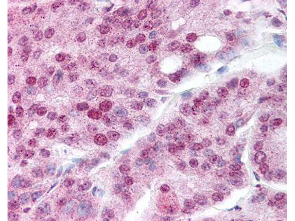
Rockland's affinity purified anti-Hsp90 acetyl K294 antibody was used at 20 μg/ml to detect signal in a variety of tissues including multi-human, multi-brain and multi-cancer slides. This image shows moderate nuclear and granular cytoplasmic positive staining in human prostate carcinoma at 40X. Tissue was formalin-fixed and paraffin embedded. The image shows localization of the antibody as the precipitated red signal, with a hematoxylin purple nuclear counterstain. Personal Communication, Tina Roush, LifeSpanBiosciences, Seattle, WA.
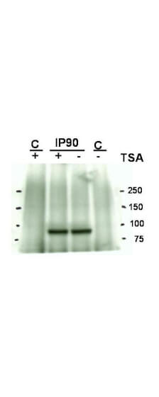
Western blot using Rockland's Affinity Purified anti-Hsp90 acetyl K294 antibody shows detection of a band at ~90 kDa corresponding to Hsp90 in an SkBr3 cell lysate (p/n W09-001-MP4). Western blotting results do not definitively demonstrate the acetyl K294 specificity of this reagent because similar staining is seen in the control lane with no treatment of TSA - Trichostatin A (an HDAC inhibitor). Immunoprecipitation with anti-Hsp90 was performed prior to western blotting with anti-Hsp90 acetyl K294.
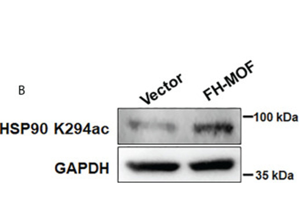
(B)?HSP90 K294 acetylation site was identified to be functioning in MOF-induced hyperacetylation of HSP90 by Western blotting assay using specific HSP90 K294ac antibody. Fig 6. PMID:?36212422
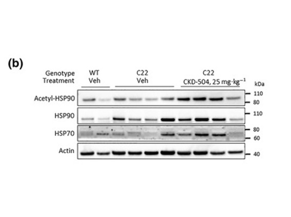
(b) The acetylation of HSP90 and expression of HSP70 were analysed (WT‐DW:?N?= 2; C22‐DW:?N?= 4; C22‐504:?N?= 4). Fig 6. PMID:?33460073
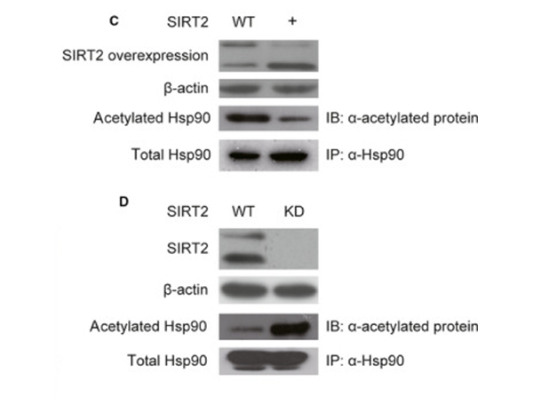
Co‐IP with anti‐Hsp90 in B104 cells overexpressing rSIRT2 (+) showed a decrease in the acetylation levels in the protein band corresponding to Hsp90 compared with control cells (WT) (C), whereas SIRT2 knock‐down?36?showed an increase in the acetylation levels of the protein band corresponding to Hsp90 compared with control cells (WT) (D). Taken together, these results suggest that Hsp90 is a direct target of SIRT2 deacetylation. Hsp90 co‐IPs (C and D) were repeated three separate times, and a representative immunoblots are shown. Fig 3. PMID:?32515550
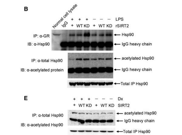
SIRT2 overexpression resulted in decreased interaction between Hsp90 and GR compared with SIRT2 knock‐down. Co‐IP assays were also performed with anti‐GR in SIRT2 overexpression, SIRT2 knocked‐down or control cells, and Hsp90 was detected in the IP complex by immunoblot (B; top panel). Co‐IP with anti‐Hsp90 under the same conditions, followed by detection of acetylated Hsp90 (B; bottom panel), showed that the interaction between Hsp90 and GR corresponds to the acetylation levels of Hsp90. (E) The B104 cell or with SIRT2 overexpression or with SIRT2 knocked‐down were treated with 10?μmol/L of Dx for 12?h. The cell lysate was incubated with anti‐Hsp90 antibody to immunoprecipitate total Hsp90 protein. Subsequently, the acetylated Hsp90 was detected in the complex through Western blot. Fig 4. PMID:?32515550
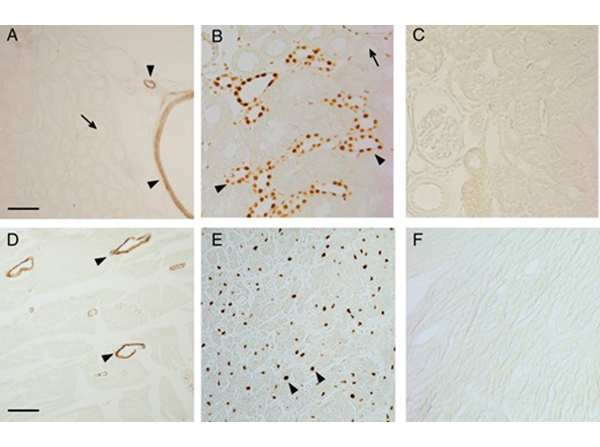
Immunohistochemical analysis of Hsp90 acetylation in rat kidney and heart shows predominant labeling in vascular smooth muscle cells. A, Representative micrograph of a paraffin-embedded section of rat kidney cortex stained with anti-Hsp90-acetyl-K295 antibody. Staining was restricted to renal arteries and arterioles (arrowheads). Renal tubules were negative for anti-Hsp90-acetyl-K295 staining (arrow). B, Rat kidney cortex stained with 12A7 anti-MR antibody. Staining is prominent in distal nephron (arrowheads) but negative in proximal tubules (arrow). C, Negative control of rat kidney cortex obtained omitting the primary anti-Hsp90-acetyl-K295 antibody. D, Representative micrograph of rat heart stained with anti-Hsp90-acetyl-K295 antibody. Staining was restricted to coronary arteries and arterioles (arrowheads). E, Rat heart stained with anti-MR antibody. Cardiomyocyte nuclei are clearly stained (arrowheads). F, Negative control of rat heart obtained omitting the primary anti-Hsp90-acetyl-K295 antibody. Bars in panels A and D, 50 μm. Fig 9. PMID:?27100623
|
|
|
|
Rockland's affinity purified anti-Hsp90 acetyl K294 antibody was used at 20 μg/ml to detect signal in a variety of tissues including multi-human, multi-brain and multi-cancer slides. This image shows moderate nuclear and granular cytoplasmic positive staining in human prostate carcinoma at 40X. Tissue was formalin-fixed and paraffin embedded. The image shows localization of the antibody as the precipitated red signal, with a hematoxylin purple nuclear counterstain. Personal Communication, Tina Roush, LifeSpanBiosciences, Seattle, WA.
|
|
| 別品名 |
rabbit anti-Heat Shock Protein HSP 90-alpha acetyl specific K294 antibody, rabbit anti-Heat Shock Protein HSP 90-alpha acetylated Lys294 antibody, HSP 86 antibody, Heat shock 86kDa antibody, Renal carcinoma antigen NY REN 38 antibody, Heat shock 90kDa protein 1 alpha antibody, Lipopolysaccharide-associated protein 2, LAP-2, HSP90A, HSPC1, HSPCA, HSP90AA1, HSP 90-α
|
| 別包装 |
あり
|
| 交差種 |
Human
|
| 適用 |
Western Blot
Enzyme Linked Immunosorbent Assay
Immunohistochemistry
|
| 免疫動物 |
Rabbit
|
| 標識物 |
Unlabeled
|
| 精製度 |
Affinity Purified
|
| 翻訳後修飾 |
アセチル化
|
| GENE ID |
3320
|
| Accession No.(Gene/Protein) |
NP_001017963.2, P07900
|
| Gene Symbol |
HSP90AA1
|
| 参考文献 |
[[Cosmo Reference Collection]][Pub Med ID]27499032,29520015,32515550,27100623,28687926,30224543,33460073,33744515,36212422,40539829
|
| [注意事項] |
濃度はロットによって異なる可能性があります。メーカーDS及びCoAからご確認ください。
|
|
| メーカー |
品番 |
包装 |
|
RKL
|
600-401-981
|
100 UG
|
※表示価格について
| 当社在庫 |
なし
|
| 納期目安 |
約10日
|
| 保存温度 |
-20℃
|
|
※当社では商品情報の適切な管理に努めておりますが、表示される法規制情報は最新でない可能性があります。
また法規制情報の表示が無いものは、必ずしも法規制に非該当であることを示すものではありません。
商品のお届け前に最新の製品法規制情報をお求めの際はこちらへお問い合わせください。
|
※当社取り扱いの試薬・機器製品および受託サービス・創薬支援サービス(納品物、解析データ等)は、研究用としてのみ販売しております。
人や動物の医療用・臨床診断用・食品用としては、使用しないように、十分ご注意ください。
法規制欄に体外診断用医薬品と記載のものは除きます。
|
|
※リンク先での文献等のダウンロードに際しましては、掲載元の規約遵守をお願いします。
|
|
※CAS Registry Numbers have not been verified by CAS and may be inaccurate.
|


