

| 別品名 |
Death up-regulated gene protein antibody, Dug antibody, H731 antibody, Ma3 antibody, Neoplastic transformation inhibitor antibody, Neoplastic transformation inhibitor protein antibody, Nuclear antigen H731 antibody
|
| 種由来 |
Human
|
| 標識物 |
Unlabeled
|
| 精製度 |
Affinity Purified
|
| 適用 |
Western Blot
Enzyme Linked Immunosorbent Assay
Immunohistochemistry
|
| 免疫動物 |
Rabbit
|
| 交差種 |
Human
|
| 翻訳後修飾 |
リン酸化
|
| GENE ID |
27250
|
| Accession No.(Gene/Protein) |
Q53EL6
|
| Gene Symbol |
PDCD4
|
| 形状 |
滅菌済み液状品
|
| 参考文献 |
[Pub Med ID]20831814
|
| [注意事項] |
濃度はロットによって異なる可能性があります。メーカーDS及びCoAからご確認ください。
|
|
※サムネイル画像をクリックすると拡大画像が表示されます。
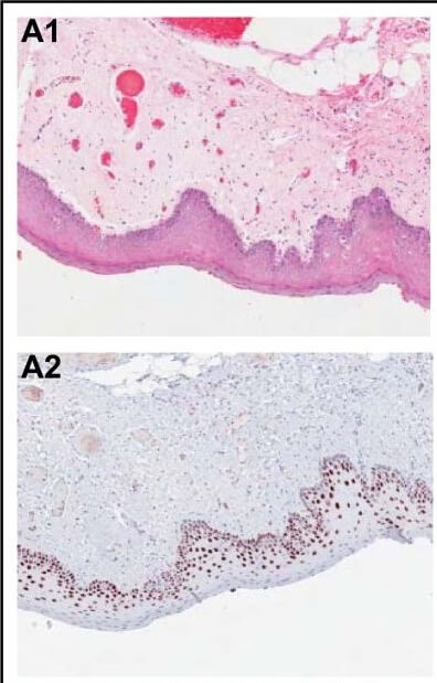
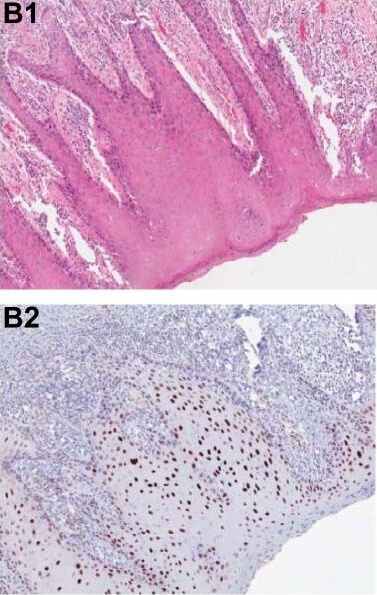
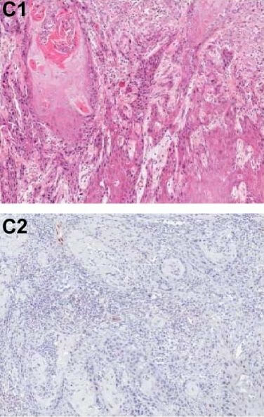
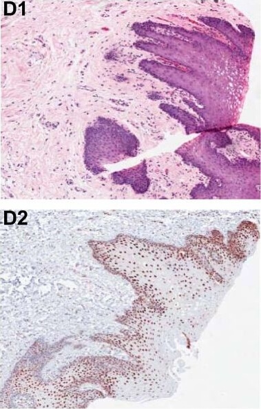
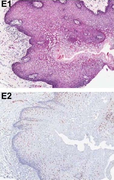
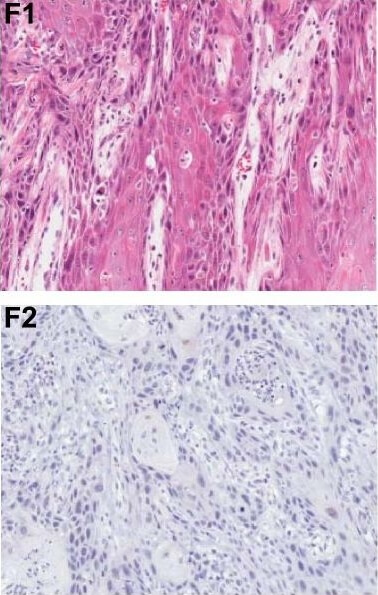
Immunohistochemical analysis of PDCD4 shows the corresponding H&E stained and PDCD4 stained tissue sections from patients with OSCC. Panels A1, A2, D1, D2 show two adjacent normal epithelium samples with strongly positive, nuclear PDCD4 staining. Panels B1, B2, E1, E2 show two dysplasia samples with positive to weak nuclear PDCD4 staining. Panels C1, C2, F1, F2 show loss of PDCD4 expression in two moderately differentiated OSCCs. Normal, dysplasia and OSCC samples are paired and correspond to two different patients (A C and D F, respectively). Figure provided by CiteAb. Source: Mol Cancer, PMID: 20831814.
Immunohistochemical analysis of PDCD4 shows the corresponding H&E-stained and PDCD4-stained tissue sections from patients with OSCC. Panels A1, A2, D1, D2 show two adjacent normal epithelium samples with strongly positive, nuclear PDCD4 staining. Panels B1, B2, E1, E2 show two dysplasia samples with positive to weak nuclear PDCD4 staining. Panels C1, C2, F1, F2 show loss of PDCD4 expression in two moderately differentiated OSCCs. Normal, dysplasia and OSCC samples are paired and correspond to two different patients (A-C and D-F, respectively). Figure provided by CiteAb. Source: Mol Cancer, PMID: 20831814.
Immunohistochemical analysis of PDCD4 shows the corresponding H&E-stained and PDCD4-stained tissue sections from patients with OSCC. Panels A1, A2, D1, D2 show two adjacent normal epithelium samples with strongly positive, nuclear PDCD4 staining. Panels B1, B2, E1, E2 show two dysplasia samples with positive to weak nuclear PDCD4 staining. Panels C1, C2, F1, F2 show loss of PDCD4 expression in two moderately differentiated OSCCs. Normal, dysplasia and OSCC samples are paired and correspond to two different patients (A-C and D-F, respectively). Figure provided by CiteAb. Source: Mol Cancer, PMID: 20831814.
Immunohistochemical analysis of PDCD4 shows the corresponding H&E-stained and PDCD4-stained tissue sections from patients with OSCC. Panels A1, A2, D1, D2 show two adjacent normal epithelium samples with strongly positive, nuclear PDCD4 staining. Panels B1, B2, E1, E2 show two dysplasia samples with positive to weak nuclear PDCD4 staining. Panels C1, C2, F1, F2 show loss of PDCD4 expression in two moderately differentiated OSCCs. Normal, dysplasia and OSCC samples are paired and correspond to two different patients (A-C and D-F, respectively). Figure provided by CiteAb. Source: Mol Cancer, PMID: 20831814.
Immunohistochemical analysis of PDCD4 shows the corresponding H&E-stained and PDCD4-stained tissue sections from patients with OSCC. Panels A1, A2, D1, D2 show two adjacent normal epithelium samples with strongly positive, nuclear PDCD4 staining. Panels B1, B2, E1, E2 show two dysplasia samples with positive to weak nuclear PDCD4 staining. Panels C1, C2, F1, F2 show loss of PDCD4 expression in two moderately differentiated OSCCs. Normal, dysplasia and OSCC samples are paired and correspond to two different patients (A-C and D-F, respectively). Figure provided by CiteAb. Source: Mol Cancer, PMID: 20831814.
Immunohistochemical analysis of PDCD4 shows the corresponding H&E-stained and PDCD4-stained tissue sections from patients with OSCC. Panels A1, A2, D1, D2 show two adjacent normal epithelium samples with strongly positive, nuclear PDCD4 staining. Panels B1, B2, E1, E2 show two dysplasia samples with positive to weak nuclear PDCD4 staining. Panels C1, C2, F1, F2 show loss of PDCD4 expression in two moderately differentiated OSCCs. Normal, dysplasia and OSCC samples are paired and correspond to two different patients (A-C and D-F, respectively). Figure provided by CiteAb. Source: Mol Cancer, PMID: 20831814.
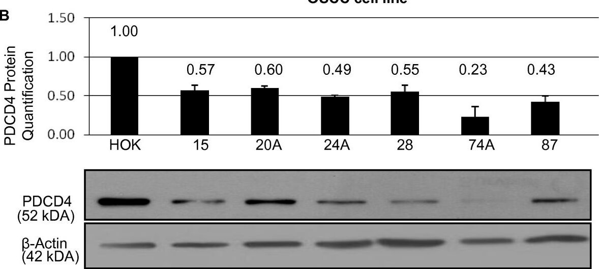
PDCD4 mRNA and PDCD4 protein levels in OSCC cell lines. (A) The log10 ratio of PDCD4 mRNA in OSCC cell lines relative to HOK. (B) Quantification of PDCD4 protein expression in OSCC cell lines with a representative Western blot of PDCD4 protein in OSCC cell lines below. Cell line data are plotted mean +- SE and are representative of 3 separate experiments. Figure provided by CiteAb. Source: Mol Cancer, PMID: 20831814.
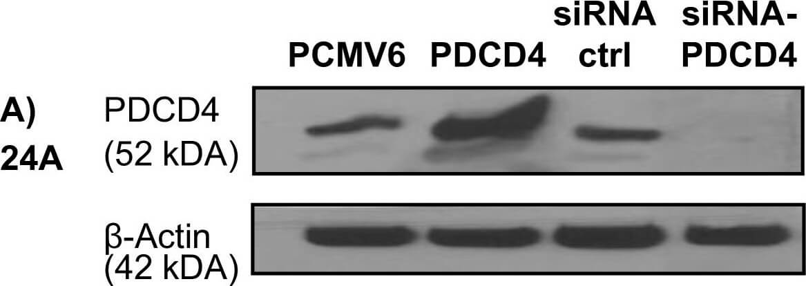
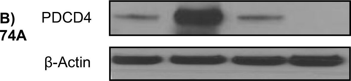
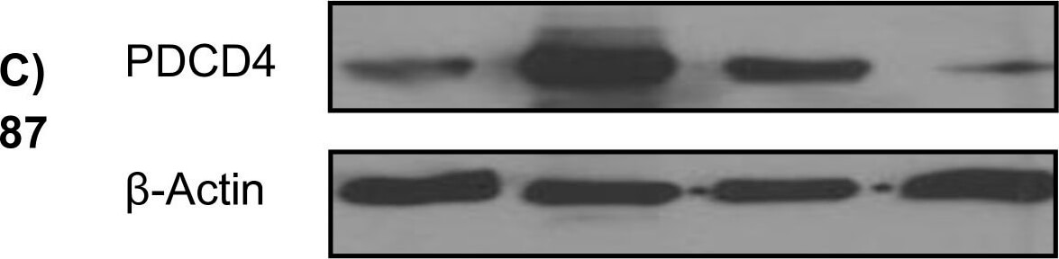
Western blotting analysis demonstrating over-expression or knock-down of PDCD4 using PDCD4 plasmid or PDCD4 targeted siRNA, respectively, versus control plasmids (PCMV6, siRNA ctrl) in the UT-SCC cell lines (A) 24A, (B) 74A, and (C) 87. Figure provided by CiteAb. Source: Mol Cancer, PMID: 20831814.
Western blotting analysis demonstrating over-expression or knock-down of PDCD4 using PDCD4 plasmid or PDCD4 targeted siRNA, respectively, versus control plasmids (PCMV6, siRNA ctrl) in the UT-SCC cell lines (A) 24A, (B) 74A, and (C) 87. Figure provided by CiteAb. Source: Mol Cancer, PMID: 20831814.
Western blotting analysis demonstrating over-expression or knock-down of PDCD4 using PDCD4 plasmid or PDCD4 targeted siRNA, respectively, versus control plasmids (PCMV6, siRNA ctrl) in the UT-SCC cell lines (A) 24A, (B) 74A, and (C) 87. Figure provided by CiteAb. Source: Mol Cancer, PMID: 20831814.
|
|
|
|
Immunohistochemical analysis of PDCD4 shows the corresponding H&E stained and PDCD4 stained tissue sections from patients with OSCC. Panels A1, A2, D1, D2 show two adjacent normal epithelium samples with strongly positive, nuclear PDCD4 staining. Panels B1, B2, E1, E2 show two dysplasia samples with positive to weak nuclear PDCD4 staining. Panels C1, C2, F1, F2 show loss of PDCD4 expression in two moderately differentiated OSCCs. Normal, dysplasia and OSCC samples are paired and correspond to two different patients (A C and D F, respectively). Figure provided by CiteAb. Source: Mol Cancer, PMID: 20831814.
|
|
|
| メーカー |
品番 |
包装 |
|
RKL
|
600-401-964
|
100 UG
|
※表示価格について
| 当社在庫 |
なし
|
| 納期目安 |
約10日
|
| 保存温度 |
-20℃
|
|

