|
※サムネイル画像をクリックすると拡大画像が表示されます。
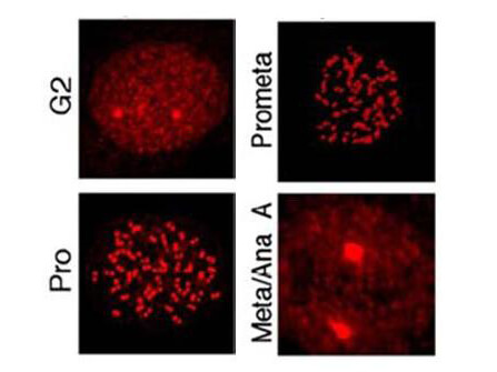
Immunostaining using Rockland's affinity purified anti-MLF1IP pT78 antibody shows detection of MLF1IP pT78 at the kinetochores of HeLa cells in different phases of the cell cycle. Fluorescent signals were detectable at the kinetochores as early as G2, became most abundant in prophase cells with a discernible nuclear envelope, and gradually diminished as cells proceeded through mitosis (Kang & Park, et al., 2006).
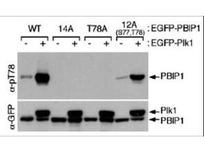
Western blot using Rockland's affinity purified anti-MLF1IP pT78 antibody shows detection of MLF1IP phosphorylated at Thr78. HeLa cells were co-infected with the indicated adenoviruses expressing GFP-tagged Plk1 or PBIP1. Blots were probed with the anti-MLF1IP pT78 antibody, stripped, and then reprobed with anti-GFP antibody (Kang & Park, et al., 2006).
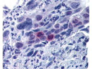
Rockland's affinity purified anti-MLF1IP pT78 antibody was used at 20 μg/ml to detect signal in a variety of tissues including multi-human, multi-brain and multi-cancer slides. This image shows moderately positive staining of mitotic cells in colon adenocarcinoma at 60X. Tissue was formalin-fixed and paraffin embedded. The image shows localization of the antibody as the precipitated red signal, with a hematoxylin purple nuclear counterstain. Personal Communication, Tina Roush, LifeSpan Biosciences, Seattle, WA.
|
|
|
|
Immunostaining using Rockland's affinity purified anti-MLF1IP pT78 antibody shows detection of MLF1IP pT78 at the kinetochores of HeLa cells in different phases of the cell cycle. Fluorescent signals were detectable at the kinetochores as early as G2, became most abundant in prophase cells with a discernible nuclear envelope, and gradually diminished as cells proceeded through mitosis (Kang & Park, et al., 2006).
|
|
| 別品名 |
rabbit anti-MLF1 pT78 antibody, rabbit anti-MLF1 Interacting Protein pT78 antibody, Centromere protein U, CENP-U, Centromere protein of 50 kDa, CENP-50, Interphase centromere complex protein 24, KSHV latent nuclear antigen-interacting protein 1, MLF1-interacting protein, Polo-box-interacting protein 1, ICEN24, PBIP1, KLIP1
|
| 交差種 |
Human
|
| 適用 |
Western Blot
Enzyme Linked Immunosorbent Assay
Immunohistochemistry
Immuno Fluorescence
|
| 免疫動物 |
Rabbit
|
| 標識物 |
Unlabeled
|
| 精製度 |
Affinity Purified
|
| 翻訳後修飾 |
リン酸化
|
| GENE ID |
79682
|
| Accession No.(Gene/Protein) |
38016935, Q71F23
|
| Gene Symbol |
CENPU
|
| 参考文献 |
Hanissian,S.H., Teng,B., Akbar,U., Janjetovic,Z., Zhou,Q., Duntsch,C. and Robertson,J.H. (2005) Regulation of myeloid leukemia factor-1 interacting protein (MLF1IP) expression in glioblastoma. Brain Res. 1047 (1), 56-64. Hanissian,S.H., Akbar,U., Teng,B., Janjetovic,Z., Hoffmann,A., Hitzler,J.K., Iscove,N., Hamre,K., Du,X., Tong,Y., Mukatira,S., Robertson,J.H. and Morris,S.W. (2004) cDNA cloning and characterization of a novel gene encoding the MLF1-interacting protein MLF1IP. Oncogene 23 (20), 3700-3707. Kang, Y.H., Park, J.E., et al. (2006) Self-Regulated Plk1 Recruitment to Kinetochores by the Plk1-PBIP1 Interaction Is Critical for Proper Chromosome Segregation. Mol. Cell 24, 409-422.
|
| [注意事項] |
濃度はロットによって異なる可能性があります。メーカーDS及びCoAからご確認ください。
|
|
| メーカー |
品番 |
包装 |
|
RKL
|
600-401-930
|
100 UG
|
※表示価格について
| 当社在庫 |
なし
|
| 納期目安 |
約10日
|
| 保存温度 |
-20℃
|
|
