

| 別品名 |
E3 ubiquitin-protein ligase Mdm2 EC=6.3.2.- p53-binding protein Mdm2 Oncoprotein Mdm2 Double minute 2 protein
|
| 抗原部位 |
a.a.177-195
|
| 種由来 |
Mouse
|
| 標識物 |
Unlabeled
|
| 精製度 |
Affinity Purified
|
| 適用 |
Western Blot
Enzyme Linked Immunosorbent Assay
|
| 免疫動物 |
Rabbit
|
| 交差種 |
Mouse
|
| GENE ID |
17246
|
| Accession No.(Gene/Protein) |
P23804
|
| Gene Symbol |
MDM2
|
| 形状 |
滅菌済み液状品
|
| 参考文献 |
Pospisilova S, Siligan C, Ban J, Jug G, Kovar H. (2004) Constitutive and DNA Damage Inducible Activation of pig3 and MDM2 Genes by Tumor-Derived p53 Mutant C277Y. Mol Cancer Res. 2(5):296-304. Aslanian A, Iaquinta PJ, Verona R, Lees JA. (2004) Repression of the Arf tumor suppressor by E2F3 is required for normal cell cycle kinetics. Genes Dev. 2004 Jun 2 [Epub ahead of print]. Stros M, Muselikova-Polanska E, Pospisilova S, Strauss F. (2004) High-Affinity Binding of Tumor-Suppressor Protein p53 and HMGB1 to Hemicatenated DNA Loops. Biochemistry. 43(22):7215-7225.
|
| [注意事項] |
濃度はロットによって異なる可能性があります。メーカーDS及びCoAからご確認ください。
|
|
※サムネイル画像をクリックすると拡大画像が表示されます。
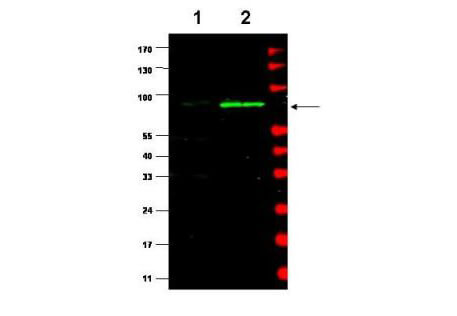
Western blot using Rockland's affinity purified Anti-MDM2 (Rabbit) is shown to detect a band (arrow) corresponding to mouse MDM2 protein. Lane 1: human kidney HEK293 cells (p/n W09-000-365). Lane 2: mouse MEF cells (p/n W10-001-371). Approximately 35μg of lysate was separated by 4-20% Tris Glycine SDS-PAGE. After blocking the membrane with 5% normal goat serum, 0.5% BLOTTO (p/n B501-0500) in PBS, the membrane was probed for overnight at 4° with the primary antibody diluted to 1:500 in 1% normal goat serum, 0.1% BLOTTO in PBS. The membrane was washed and reacted with a 1:10,000 dilution of IRDye800 conjugated Gt-a-Rabbit IgG [H&L] (p/n 611-132-122) for 45 min at room temperature (800 nm channel, green). Molecular weight estimation was made by comparison to prestained MW markers indicated at the right (700 nm channel, red). IRDye800 fluorescence image was captured using the OdysseyR Infrared Imaging System developed by LI-COR. IRDye is a trademark of LI-COR, Inc. Other detection systems will yield similar results.
|
|
|
|
Western blot using Rockland's affinity purified Anti-MDM2 (Rabbit) is shown to detect a band (arrow) corresponding to mouse MDM2 protein. Lane 1: human kidney HEK293 cells (p/n W09-000-365). Lane 2: mouse MEF cells (p/n W10-001-371). Approximately 35μg of lysate was separated by 4-20% Tris Glycine SDS-PAGE. After blocking the membrane with 5% normal goat serum, 0.5% BLOTTO (p/n B501-0500) in PBS, the membrane was probed for overnight at 4° with the primary antibody diluted to 1:500 in 1% normal goat serum, 0.1% BLOTTO in PBS. The membrane was washed and reacted with a 1:10,000 dilution of IRDye800 conjugated Gt-a-Rabbit IgG [H&L] (p/n 611-132-122) for 45 min at room temperature (800 nm channel, green). Molecular weight estimation was made by comparison to prestained MW markers indicated at the right (700 nm channel, red). IRDye800 fluorescence image was captured using the OdysseyR Infrared Imaging System developed by LI-COR. IRDye is a trademark of LI-COR, Inc. Other detection systems will yield similar results.
|
|
|
| メーカー |
品番 |
包装 |
|
RKL
|
600-401-927
|
100 UG
|
※表示価格について
| 当社在庫 |
なし
|
| 納期目安 |
約10日
|
| 保存温度 |
-20℃
|
|

