|
※サムネイル画像をクリックすると拡大画像が表示されます。
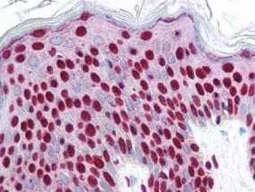
Rockland's affinity purified anti-Smad3 pS423 pS425 antibody was used at 2.5 ug/ml to detect signal in a variety of tissues including multi-human, multi-brain and multi-cancer slides. This image shows strong nuclear staining in the majority of epidermal keratinocytes at 40X. Tissue was formalin-fixed and paraffin embedded. The image shows localization of the antibody as the precipitated red signal, with a hematoxylin purple nuclear counterstain. Personal Communi-cation, Tina Roush, LifeSpanBiosciences, Seattle, WA.
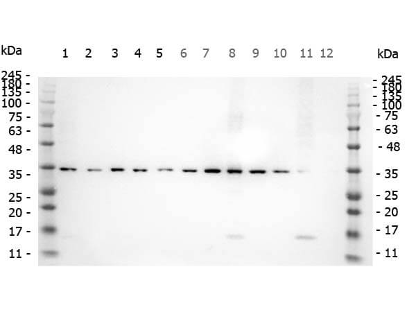
Western Blot of Rabbit anti-SMAD3 pS423 pS425 antibody. Marker: Opal Pre-stained ladder (p/n MB-210-0500). Lane 1: HEK293 lysate (p/n W09-000-365). Lane 2: HeLa Lysate (p/n W09-000-364). Lane 3: MCF-7 Lysate (p/n W09-000-360). Lane 4: Jurkat Lysate (p/n W09-000-370). Lane 5: A431 Lysate (p/n W09-000-361). Lane 6: A549 Lysate (p/n W09-001-372). Lane 7: LNCap Lysate (p/n W09-001-GJ9). Lane 8: MOLT-4 Lysate (p/n W09-001-GK2). Lane 9: Ramos Lysate (p/n W09-000-GK4). Lane 10: Raji Lysate (p/n W09-001-368). Lane 11: A-172 Lysate (p/n W09-001-GL5). Lane 12: NIH/3T3 Lysate (p/n W10-000-358). Load: 10 μg per lane. Primary antibody: SMAD3 pS423 pS425antibody at 1ug/mL overnight at 4C. Secondary antibody: Peroxidase rabbit secondary antibody (p/n 611-103-122) at 1:30,000 for 60 min at RT. Blocking Buffer: 1% Casein-TTBS (p/n MB-082) for 30 min at RT. Predicted/Observed size: 35 kDa for SMAD3 pS423 pS425.
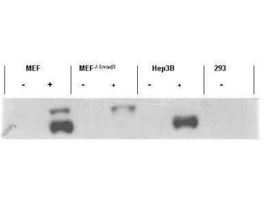
Western blot using Rockland's affinity purified anti-Smad3 pS423 pS425 antibody shows detection of endogenous Smad3 in stimulated cell lysates. Lysates were prepared from control cells (- lanes), or cells stimulated with 2 ng/ml TGF (+lanes) for 1 hour. This reagent recognizes phosphorylated Smad3 and has negligible reactivity against non-phosphorylated Smad3 protein. Personal Communication. Ying Zhang, NIH, CCR, Bethesda, MD.
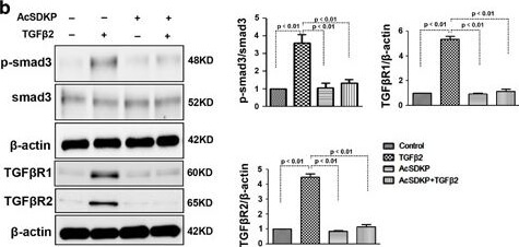
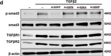
Proximity between AcSDKP and FGFR1 inhibits the TGFβ/smad signaling pathway in HMVECs. (a) HMVECs were treated with N-FGFR1 (1.5??μg/ml) for 48?h with or without preincubation with AcSDKP (100?nM) for 2?h, and the proximity between AcSDKP and FGFR1 was analyzed by the Duolink In Situ Assay. For each slide, images at a × 400 original magnification were obtained from six different areas. (b and c) HMVECs were treated with TGFβ2 (5?ng/ml) for 15?min or 48?h with or without preincubation with AcSDKP for 2?h, and the p-smad3, TGFβR1, TGFβR2 and FGFR1 levels were analyzed by western blot. Densitometric analysis of the p-smad3/smad3, TGFβR1/β-actin, TGFβR2/β-actin and FGFR1/β-actin levels from each group (n=6) were analyzed. (d and e) HMVECs were incubated with TGFβ2 for 15?min or 48?h with or without preincubation with AcSDKP or its mutants (AcDSPK, AcSDKA, AcADKP) (100?nM) for 2?h. The p-smad3/smad3, TGFβR1/β-actin, TGFβR2/β-actin and FGFR1/β-actin protein levels were analyzed by western blot Figure provided by CiteAb. Source: Cell Death Dis, PMID: 28771231.
Proximity between AcSDKP and FGFR1 inhibits the TGFβ/smad signaling pathway in HMVECs. (a) HMVECs were treated with N-FGFR1 (1.5??μg/ml) for 48?h with or without preincubation with AcSDKP (100?nM) for 2?h, and the proximity between AcSDKP and FGFR1 was analyzed by the Duolink In Situ Assay. For each slide, images at a × 400 original magnification were obtained from six different areas. (b and c) HMVECs were treated with TGFβ2 (5?ng/ml) for 15?min or 48?h with or without preincubation with AcSDKP for 2?h, and the p-smad3, TGFβR1, TGFβR2 and FGFR1 levels were analyzed by western blot. Densitometric analysis of the p-smad3/smad3, TGFβR1/β-actin, TGFβR2/β-actin and FGFR1/β-actin levels from each group (n=6) were analyzed. (d and e) HMVECs were incubated with TGFβ2 for 15?min or 48?h with or without preincubation with AcSDKP or its mutants (AcDSPK, AcSDKA, AcADKP) (100?nM) for 2?h. The p-smad3/smad3, TGFβR1/β-actin, TGFβR2/β-actin and FGFR1/β-actin protein levels were analyzed by western blot Figure provided by CiteAb. Source: Cell Death Dis, PMID: 28771231.
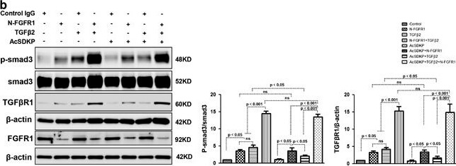
AcSDKP suppresses TGFβ/smad signaling and EndMT through the FGFR1/FRS2 pathway. (a) HMVECs were treated with N-FGFR1 for 48?h, and the FGFR1, TGFβR1 and TGFβR2 protein levels were analyzed by western blot. (b) HMVECs were treated with TGFβ2 in the presence or absence of N-FGFR1 for 15?min with or without AcSDKP preincubation. The p-smad3 and TGFβR1 protein levels were analyzed by western blot. Densitometric analysis of the p-smad3/smad3 and TGFβR1/β-actin levels (n=3) in each group was performed. (c) HMVECs were incubated with either N-FGFR1 in the presence or absence of TGFβ2 for 48?h with or without preincubation with AcSDKP for 2?h or with N-FGFR1 in the presence or absence of TGFβ2 for 48?h with or without 24?h of incubation with FGF2 (50?ng/ml). The CD31, SM22α, FSP1 and α-SMA protein levels were analyzed by western blot. (d) HMVECs were transfected with FRS2 siRNA (100?nM) for 48?h with or without AcSDKP preincubation. The VE-cadherin, FSP1, vimentin, SM22α and p-smad3 levels were analyzed by western blot. (e) HMVECs were treated with N-FGFR1 for 48?h or 15?min in the presence or absence of N-TGFβ (1, 2, 3) (1.0??μg/ml). The CD31, VE-cadherin, SM22α, FSP1, TGFβR1, TGFβR2 and p-smad3 levels were analyzed by western blot Figure provided by CiteAb. Source: Cell Death Dis, PMID: 28771231.
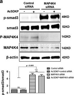
MAP4K4 deficiency induces TGFβ/smad signaling and EndMT via activation of integrin β1. (a) HMVECs were transfected with MAP4K4 siRNA (100?nM) for 48?h. Next, the cells were treated with or without AcSDKP for 2?h. The p-smad3/smad3 pathway was analyzed by western blot. Densitometric analysis of the p-smad3/smad3 levels was performed, with n=3 for each group. (b) HMVECs were treated with MAP4K4 siRNA for 48?h with or without AcSDKP treatment. The VE-cadherin, CD31, FSP1, SM22α and vimentin protein levels were analyzed by western blot. (c) HMVECs were transfected with MAP4K4 siRNA for 48?h in the presence or absence of TGFβ2 with or without AcSDKP. The integrin β1 level was analyzed by western blot Figure provided by CiteAb. Source: Cell Death Dis, PMID: 28771231.
|

|
|
Rockland's affinity purified anti-Smad3 pS423 pS425 antibody was used at 2.5 ug/ml to detect signal in a variety of tissues including multi-human, multi-brain and multi-cancer slides. This image shows strong nuclear staining in the majority of epidermal keratinocytes at 40X. Tissue was formalin-fixed and paraffin embedded. The image shows localization of the antibody as the precipitated red signal, with a hematoxylin purple nuclear counterstain. Personal Communi-cation, Tina Roush, LifeSpanBiosciences, Seattle, WA.
|
|
| 別品名 |
rabbit anti-SMAD3 pS423pS425 antibody, SMAD-3, SMAD 3, mothers against decapentaplegic homolog 3 antibody, MAD homolog 3, Mothers against DPP homolog 3, SMAD family member 3, MADH3, MADH 3, JV15-2, nothing
|
| 交差種 |
Human
|
| 適用 |
Western Blot
Enzyme Linked Immunosorbent Assay
Immunohistochemistry
|
| 免疫動物 |
Rabbit
|
| 抗原部位 |
a.a.417-425
|
| 標識物 |
Unlabeled
|
| 精製度 |
Affinity Purified
|
| 翻訳後修飾 |
リン酸化
|
| GENE ID |
4088
|
| Accession No.(Gene/Protein) |
5174513, P84022
|
| Gene Symbol |
SMAD3
|
| 参考文献 |
[Pub Med ID]18922473
|
| [注意事項] |
濃度はロットによって異なる可能性があります。メーカーDS及びCoAからご確認ください。
|
|
| メーカー |
品番 |
包装 |
|
RKL
|
600-401-919
|
100 UG
|
※表示価格について
| 当社在庫 |
なし
|
| 納期目安 |
約10日
|
| 保存温度 |
-20℃
|
|


