|
※サムネイル画像をクリックすると拡大画像が表示されます。
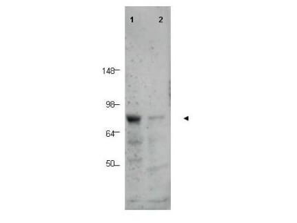
Western blot using Rockland's Affinity Purified anti-BORIS antibody shows detection of a predominant band corresponding to BORIS in human tissue lysates (arrowhead).?? Lane 1 contains lysate from human prostate tissue.? Lane 2 contains lysate from human spleen tissue.? A predominant band at ~75 kDa is observed.? Molecular weight estimation was made by comparison to prestained MW markers as indicated.
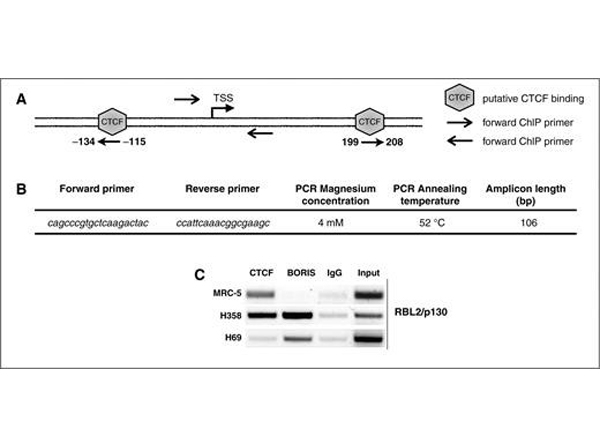
A, schematic representation of?Rb2/p130?gene region containing CTCF putative binding sites. B, nucleotide sequence of the primers spanning the?Rb2/p130?region investigated. C,?in vivo?binding (XChIP) of CTCF and Boris to?Rb2/p130?(RBL2/P130) promoter in MRC-5 lung fibroblasts, and H358 (NCSCL) and H69 (SCLC)lung cancer cell lines. The XChIPs were performed using the ChIP-IT Express Enzymatic kit (Active Motif). For each immunoprecipitation, 10 μg of sheared chromatin was incubated overnight with 2 μg of polyclonal antibody specific for CTCF (Millipore, DAM1421463), BORIS (Rockland, 600?401-907) or normal rabbit IgG (Santa Cruz, sc-2027). PCR was then performed on 1/10 of immunoprecipitated DNA using specific primers for Rb2/p130 core promoter region (B). As positive control for CTCF and BORIS DNA?protein binding, PCR was performed amplyfing the?H19/DMR?locus (Nguyen, 2008; ref.?21). Input represents the 0.25% of total preimmunoprecipitated chromatin. The results were confirmed by three independent experiments. Fig 1. PMID:?21325284
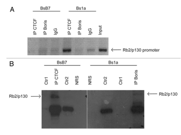
A) In vivo binding (XChIP) of CTCF and BORIS to Rb2/p130 promoter in BsB7 (T-Ag-positive) and Bs1a (T-Ag-negative) mouse medulloblastoma cell lines. For each immunoprecipitation, 10 μg of sheared chromatin was incubated overnight with 2 μg of polyclonal antibody specific for CTCF (Millipore, DAM1421463), BORIS (Rockland, 600-401-907) or normal rabbit IgG (Santa Cruz, sc-2027). PCR was then performed on 1/10 of immunoprecipitated DNA using specific primers for Rb2/p130 core promoter region (Fiorentino et al., 2011). As positive control for CTCF and CTCFL/BORIS DNA-protein binding, PCR was performed by amplifying the H19/DMR locus (21). Input represents 0.25% of total preimmunoprecipitated chromatin. The results were confirmed by three independent experiments; B) Rb2/p130 co-immunoprecipitates with CTCF and BORIS in mouse medulloblastoma BsB7 (T-Ag-positive) cells. Immunoprecipitation experiments were performed from total fractions and using rabbit polyclonal anti-CTCF or anti-BORIS as immunoprecipitating antibodies. The presence of Rb2/p130 in both CTCF and BORIS precipitates was assessed with mouse monoclonal anti-Rb2/p130 antibody by western blotting. Ctr1 (Supernatant+Antibody) and Ctr2 (Beads+Antibody) were used as immunoprecipitation controls. Figure?2.?PMID:?22544282
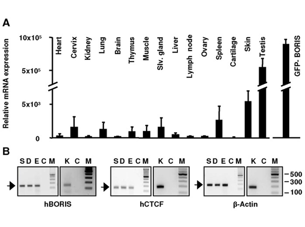
Expression of BORIS and CTCF in mouse tissues and human skin. A) BORIS mRNA expression in mouse tissues as analysed by quantitative RT-PCR by the comparative Ct method and normalised to GAPDH. Data are represented as fold changes relative to the lowest BORIS/GAPDH ratio (cartilage, designated as 1.0). For each sample, measurements were done in duplicate using two different primer sets. Error bars represent s.d. HEK293T cells transfected with pEGFP-mBORIS were used as positive control (right graph).?B) BORIS, CTCF and β-Actin (internal control) mRNA expression in human skin and primary keratinocytes by semiquantitative RT-PCR (H0.3 primer set was used, see?Information S1). Human total skin (S), dermis (D), epidermis (E) and freshly isolated keratinocytes (K), buffer only-control (C) or molecular weight markers (M).?Figure 1. PMID:?22724006
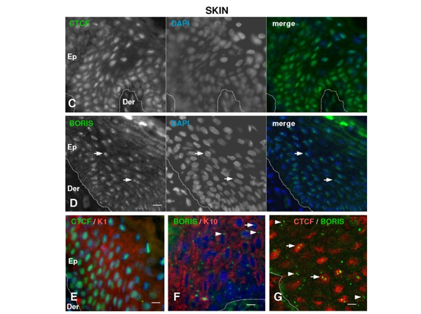
Expression of BORIS and CTCF in human skin. C,D) Indirect immunofluorescence experiments on human skin sections with anti-CTCF or anti-BORIS antibodies as indicated. Colours as indicated. The nuclei were visualised with DAPI (blue). Dotted line indicates the basal membrane that separates the epidermis (Ep) from the dermis (Der). Scale bar: 50 μm. Photographs are representative of studies on five different human individuals with three different polyclonal antibodies for BORIS and two different antibodies for CTCF.?E,F) Double immunofluorescence for CTCF or BORIS and markers of post-mitotic terminal differentiation keratins K1/K10. Colours as indicated. Scale bar: 20 μm.?G) Double immunofluorescence for CTCF (red) or BORIS (green). Scale bar: 10 μm. Arrows indicate the focal accumulation of BORIS within the nuclei, arrowheads indicate BORIS dots beside the nuclei. Colours as indicated. Figure 1. PMID:?22724006
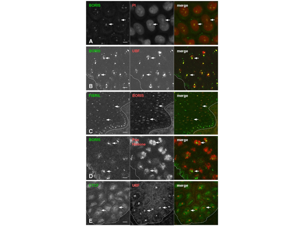
BORIS localises to the nucleoli of human epidermal cells. A?E). Double immunofluorescence analyses were performed on human skin sections with antibodies to BORIS or CTCF and UBF, Fibrillarin or pan-histone, as indicated. Coulors as indicated. Nucleoli were counterstained with propidium iodie (PI) in A. Arrows point at nucleoli (A?C,E) or histone-dark areas (D). Note the coincidence between the BORIS protein, nucleolar markers and dark-histone areas. Scale bar: 20 μm. Fig 2. PMID:?22724006
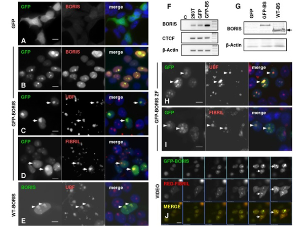
Exogenous GFP-BORIS localises to the nucleoli. A?E, H,I. Detection of GFP (green) or BORIS (red), UBF, or Fibrillarin by immunofluorescence in HEK293T cells 24 hours after transfection with plasmids carrying GFP (A) or GFP-BORIS (B-D), wild-type BORIS (E) or GFP-Zinc finger domain of BORIS (H,I), as indicated on the left side. The nuclei were visualised with DAPI (blue). Scale bar: 10 μm.?F) Detection of BORIS mRNA expression by RT-PCR in HEK293T cells transiently transfected as above. Primers for CTCF and β-actin were used as controls. BS: BORIS.?G) Detection of BORIS by western blotting with the antibodies used for the immunofluorescence analyses above. Note the higher molecular weight of the fusion protein GFP-BORIS, compared to the wild-type protein (arrow). β-Actin as loading control.?J) Video microphotograms showing the co-localisation of GFP-BORIS and Fibrillarin-Cherry in live cells after transient transfection (see also?Video S1). Fig 3. PMID:?22724006
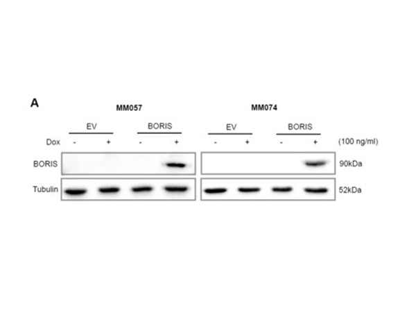
A) Expression of BORIS fused to a triple FLAG-tag was induced in the MM057 and MM074 melanoma cell lines with 100?ng/ml dox for 5 days.??Whole-cell lysate was used for immunoblotting (n?=?3) with anti-BORIS antibody. Anti-Tubulin was used as a loading control. Fig 6. PMID:?32123577
|
|
|
|
Western blot using Rockland's Affinity Purified anti-BORIS antibody shows detection of a predominant band corresponding to BORIS in human tissue lysates (arrowhead).?? Lane 1 contains lysate from human prostate tissue.? Lane 2 contains lysate from human spleen tissue.? A predominant band at ~75 kDa is observed.? Molecular weight estimation was made by comparison to prestained MW markers as indicated.
|
|
| 別品名 |
rabbit anti-BORIS Antibody, Transcriptional repressor CTCFL, Brother of the regulator of imprinted sites antibody, CCCTC binding factor (zinc finger protein) like antibody, CCCTC-binding factor antibody, CTCF paralog antibody, CTCF-like, cancer/testis antigen 27, CT27, CTCF-T
|
| 交差種 |
Human
|
| 適用 |
Western Blot
Enzyme Linked Immunosorbent Assay
|
| 免疫動物 |
Rabbit
|
| 抗原部位 |
a.a.9-26
|
| 標識物 |
Unlabeled
|
| 精製度 |
Affinity Purified
|
| GENE ID |
140690
|
| Accession No.(Gene/Protein) |
20805280, Q8NI51
|
| Gene Symbol |
CTCFL
|
| 参考文献 |
[[Cosmo Reference Collection]][Pub Med ID]18355444,20305816,21325284,22544282,22724006,32123577
|
| [注意事項] |
濃度はロットによって異なる可能性があります。メーカーDS及びCoAからご確認ください。
|
|
| メーカー |
品番 |
包装 |
|
RKL
|
600-401-907
|
100 UG
|
※表示価格について
| 当社在庫 |
なし
|
| 納期目安 |
約10日
|
| 保存温度 |
-20℃
|
|


