
| 別品名 |
Epidermal growth factor receptor antibody, erbb 1 antibody, Erbb antibody, Erbb1 antibody, HER1 antibody, mENA antibody
|
| 抗原部位 |
a.a.1189-1199
|
| 種由来 |
Human
|
| 標識物 |
Unlabeled
|
| 精製度 |
Serum
|
| 適用 |
Western Blot
Enzyme Linked Immunosorbent Assay
|
| 免疫動物 |
Rabbit
|
| 交差種 |
Human
|
| GENE ID |
1956
|
| Accession No.(Gene/Protein) |
P00533
|
| Gene Symbol |
EGFR
|
| 形状 |
滅菌済み液状品
|
| 参考文献 |
[Pub Med ID]29357089, 30641288, 27260220
|
| [注意事項] |
濃度はロットによって異なる可能性があります。メーカーDS及びCoAからご確認ください。
|
|
※サムネイル画像をクリックすると拡大画像が表示されます。
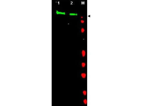
Western blot using Rockland's Affinity Purified anti EGFR antibody shows detection of a band at ~170 kDa corresponding to human EGFR (arrowhead). Lane 1: unstimulated A431 whole cell lysate (p/n W09 000 361). Lane 2: EGF (50 ng/ml for 15 min) stimulated A431 whole cell lysates (p/n W09 000 362). Approximately 30ug of lysate was separated on a 4 20% Tris Glycine gel by SDS PAGE and transferred onto nitrocellulose. After blocking the membrane was probed with the primary antibody diluted to 1:1,000. Reaction occurred overnight at 4 C followed by washes and reaction with a 1:10,000 dilution of IRDye800 conjugated Gt a Rabbit IgG [H&L] MX (p/n 611 132 122) for 45 min at room temperature (800 nm channel, green). Molecular weight estimation was made by comparison to prestained MW markers in lane M (700 nm channel, red). IRDye800 fluorescence image was captured using the OdysseyR Infrared Imaging System developed by LI COR. IRDye is a trademark of LI COR, Inc. Other detection systems will yield similar results.
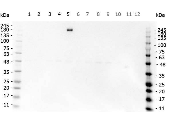
Western Blot of Rabbit anti-EGFR antibody. Marker: Opal Pre-stained ladder (p/n MB-210-0500). Lane 1: HEK293 lysate (p/n W09-000-365). Lane 2: HeLa Lysate (p/n W09-000-364). Lane 3: MCF-7 Lysate (p/n W09-000-360). Lane 4: Jurkat Lysate (p/n W09-000-370). Lane 5: A431 Lysate (p/n W09-000-361). Lane 6: A549 Lysate (p/n W09-001-372). Lane 7: LNCap Lysate (p/n W09-001-GJ9). Lane 8: MOLT-4 Lysate (p/n W09-001-GK2). Lane 9: Ramos Lysate (p/n W09-000-GK4). Lane 10: Raji Lysate (p/n W09-001-368). Lane 11: A-172 Lysate (p/n W09-001-GL5). Lane 12: NIH/3T3 Lysate (p/n W10-000-358). Load: 35 ug per lane. Primary antibody: EGFR antibody at 1ug/mL overnight at 4C. Secondary antibody: Peroxidase rabbit secondary antibody (p/n 611-103-122) at 1:30,000 for 60 min at RT. Blocking Buffer: 1% Casein-TTBS (p/n MB-082) for 30 min at RT. Predicted/Observed size: 170kDa for EGFR.
|
|
|
|
Western blot using Rockland's Affinity Purified anti EGFR antibody shows detection of a band at ~170 kDa corresponding to human EGFR (arrowhead). Lane 1: unstimulated A431 whole cell lysate (p/n W09 000 361). Lane 2: EGF (50 ng/ml for 15 min) stimulated A431 whole cell lysates (p/n W09 000 362). Approximately 30ug of lysate was separated on a 4 20% Tris Glycine gel by SDS PAGE and transferred onto nitrocellulose. After blocking the membrane was probed with the primary antibody diluted to 1:1,000. Reaction occurred overnight at 4 C followed by washes and reaction with a 1:10,000 dilution of IRDye800 conjugated Gt a Rabbit IgG [H&L] MX (p/n 611 132 122) for 45 min at room temperature (800 nm channel, green). Molecular weight estimation was made by comparison to prestained MW markers in lane M (700 nm channel, red). IRDye800 fluorescence image was captured using the OdysseyR Infrared Imaging System developed by LI COR. IRDye is a trademark of LI COR, Inc. Other detection systems will yield similar results.
|
|
|
| メーカー |
品番 |
包装 |
|
RKL
|
600-401-905
|
100 UG
|
※表示価格について
| 当社在庫 |
なし
|
| 納期目安 |
約10日
|
| 保存温度 |
-20℃
|
|

