|
※サムネイル画像をクリックすると拡大画像が表示されます。
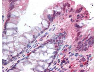
Rockland's affinity purified anti-NOXO1 antibody was used at 5 ug/ml to detect signal in a variety of tissues including multi-human, multi-brain and multi-cancer slides. This image shows moderate positive staining of the lamina propia in human colon epithelium and macrophages at 40X. Tissue was formalin-fixed and paraffin embedded. The image shows localization of the antibody as the precipitated red signal, with a hematoxylin purple nuclear counterstain. Personal Communication, Tina Roush, LifeSpan Biosciences, Seattle, WA.
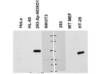
Western blot using Rockland's Affinity Purified anti-NOXO1 antibody shows detection of a band ~50 kDa corresponding to human NOXO1. Reactivity was observed in transfected human 293 cells and human HT-29 colon carcinoma cells (endogenous). Under these conditions endogenous NOXO1 detection was not observed in HeLa, HL-60, 3T3, untransfected 293 or WT MEF cells. A 1:1,000 dilution of the primary antibody was used for detection followed by secondary antibody reactivity. Specific band reactivity was competed away when the antibody was pre-incubated with the peptide immunogen (data not shown). Personal Communication, Zhenggang Liu, NIH, CCR, Bethesda, MD.
|
|
|
|
Rockland's affinity purified anti-NOXO1 antibody was used at 5 ug/ml to detect signal in a variety of tissues including multi-human, multi-brain and multi-cancer slides. This image shows moderate positive staining of the lamina propia in human colon epithelium and macrophages at 40X. Tissue was formalin-fixed and paraffin embedded. The image shows localization of the antibody as the precipitated red signal, with a hematoxylin purple nuclear counterstain. Personal Communication, Tina Roush, LifeSpan Biosciences, Seattle, WA.
|
|
| 別品名 |
rabbit anti-NOXO1 antibody, NOXO-1, NOXO 1, NADPH oxidase organizer 1 antibody, NADPH oxidase regulatory protein antibody, Nox organizer 1 antibody, Nox organizing protein 1 antibody, Regulatory protein P41NOX antibody, SH3 and PX domain-containing protein 5, P41NOX, SH3PXD5
|
| 交差種 |
Human
|
| 適用 |
Western Blot
Enzyme Linked Immunosorbent Assay
Immunohistochemistry
|
| 免疫動物 |
Rabbit
|
| 抗原部位 |
a.a.238-252
|
| 標識物 |
Unlabeled
|
| 精製度 |
Affinity Purified
|
| GENE ID |
124056
|
| Accession No.(Gene/Protein) |
16198473, Q8NFA2
|
| Gene Symbol |
NOXO1
|
| 参考文献 |
[Pub Med ID]34127487
|
| [注意事項] |
濃度はロットによって異なる可能性があります。メーカーDS及びCoAからご確認ください。
|
|
| メーカー |
品番 |
包装 |
|
RKL
|
600-401-899
|
100 UG
|
※表示価格について
| 当社在庫 |
なし
|
| 納期目安 |
約10日
|
| 保存温度 |
-20℃
|
|
※当社では商品情報の適切な管理に努めておりますが、表示される法規制情報は最新でない可能性があります。
また法規制情報の表示が無いものは、必ずしも法規制に非該当であることを示すものではありません。
商品のお届け前に最新の製品法規制情報をお求めの際はこちらへお問い合わせください。
|
※当社取り扱いの試薬・機器製品および受託サービス・創薬支援サービス(納品物、解析データ等)は、研究用としてのみ販売しております。
人や動物の医療用・臨床診断用・食品用としては、使用しないように、十分ご注意ください。
法規制欄に体外診断用医薬品と記載のものは除きます。
|
|
※リンク先での文献等のダウンロードに際しましては、掲載元の規約遵守をお願いします。
|
|
※CAS Registry Numbers have not been verified by CAS and may be inaccurate.
|
