|
※サムネイル画像をクリックすると拡大画像が表示されます。
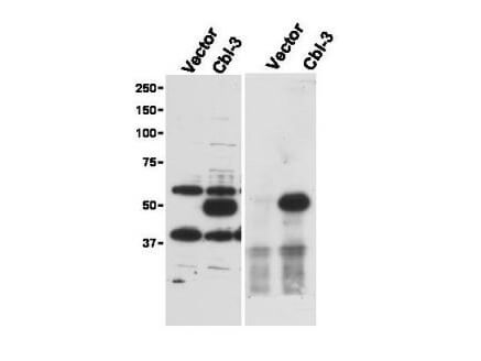
Immunoprecipitation (right) and western blot (left) using Rockland's Affinity Purified anti-Cbl-c antibody shows detection of a predominant band at ~52 kDa corresponding to Cbl-c. Lysates used are from HEK293T cells transfected with empty vector or with Cbl-c and western blotting (left panel). The predicted size of Cbl-c is 52 kDa. Size markers in kDa are shown to the left of the panel. The (right panel) shows immunoprecipitation with Rabbit anti-Cbl-c followed by western blotting using a Goat anti-Cbl-c antibody. Personal Communication. Stan Lipkowitz, NCI, NIH, Bethesda, MD.
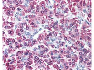
Rockland's affinity purified anti-Cbl-c antibody was used at 5 μg/ml to detect signal in a variety of tissues including multi-human, multi-brain and multi-cancer slides. This image shows moderate intracellular positive staining of human pancreatic acinar epithelium at 40X. Tissue was formalin-fixed and paraffin embedded. The image shows localization of the antibody as the precipitated red signal, with a hematoxylin purple nuclear counterstain. Personal Communication, Tina Roush, LifeSpanBiosciences, Seattle, WA.
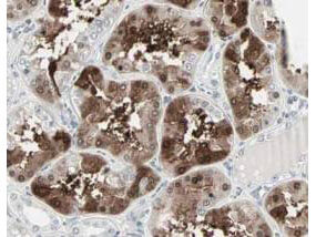
Rockland's Affinity Purified anti-Cbl-c antibody shows strong nuclear and cytoplasmic staining of cells in tubuli in human kidney tissue. Tissue was formalin-fixed and paraffin embedded. Brown color indicates presence of protein, blue color shows cell nuclei. Personal Communication, Kenneth Wester, www.proteinatlas.org, Uppsala, Sweden.
|
|
|
|
Immunoprecipitation (right) and western blot (left) using Rockland's Affinity Purified anti-Cbl-c antibody shows detection of a predominant band at ~52 kDa corresponding to Cbl-c. Lysates used are from HEK293T cells transfected with empty vector or with Cbl-c and western blotting (left panel). The predicted size of Cbl-c is 52 kDa. Size markers in kDa are shown to the left of the panel. The (right panel) shows immunoprecipitation with Rabbit anti-Cbl-c followed by western blotting using a Goat anti-Cbl-c antibody. Personal Communication. Stan Lipkowitz, NCI, NIH, Bethesda, MD.
|
|
| 別品名 |
rabbit anti-Cbl-c Antibody, E3 ubiquitin-protein ligase CBL-C, RING finger protein 57, RING-type E3 ubiquitin transferase CBL-C, SH3-binding protein CBL-3, SH3-binding protein CBL-C, Signal transduction protein CBL-C, CBLC, CBL3, RNF57
|
| 交差種 |
Human
|
| 適用 |
Western Blot
Enzyme Linked Immunosorbent Assay
Immunohistochemistry
Immunoprecipitation
|
| 免疫動物 |
Rabbit
|
| 抗原部位 |
a.a.444-458
|
| 標識物 |
Unlabeled
|
| 精製度 |
Affinity Purified
|
| GENE ID |
23624
|
| Accession No.(Gene/Protein) |
125987803, Q9ULV8
|
| Gene Symbol |
CBLC
|
| 参考文献 |
[Pub Med ID]29945960
|
| [注意事項] |
濃度はロットによって異なる可能性があります。メーカーDS及びCoAからご確認ください。
|
|
| メーカー |
品番 |
包装 |
|
RKL
|
600-401-889
|
100 UG
|
※表示価格について
| 販売状況 |
長期B.O、納期未定
|
| 当社在庫 |
なし
|
| 納期目安 |
納期未定
|
| 保存温度 |
-20℃
|
|
※当社では商品情報の適切な管理に努めておりますが、表示される法規制情報は最新でない可能性があります。
また法規制情報の表示が無いものは、必ずしも法規制に非該当であることを示すものではありません。
商品のお届け前に最新の製品法規制情報をお求めの際はこちらへお問い合わせください。
|
※当社取り扱いの試薬・機器製品および受託サービス・創薬支援サービス(納品物、解析データ等)は、研究用としてのみ販売しております。
人や動物の医療用・臨床診断用・食品用としては、使用しないように、十分ご注意ください。
法規制欄に体外診断用医薬品と記載のものは除きます。
|
|
※リンク先での文献等のダウンロードに際しましては、掲載元の規約遵守をお願いします。
|
|
※CAS Registry Numbers have not been verified by CAS and may be inaccurate.
|
