|
※サムネイル画像をクリックすると拡大画像が表示されます。
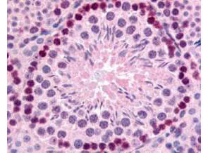
Rockland's Affinity Purified anti-mouse Gli-2 antibody was used at 10 μg/ml to evaluate staining on several mouse tissues. Moderate to strong staining was seen on many tissues, with low background staining. This image shows Gli-2 staining of mouse testis. Tissue was formalin-fixed and paraffin embedded.
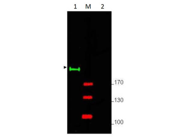
Western blot using Rockland's Affinity Purified anti-Gli-2 antibody shows detection of a predominant band at ~190 kDa corresponding to Gli-2 (arrowhead) in mouse brain whole cell lysate (p/n W10-000-T004) (lane 1). Pre-incubation of antibody with immunizing peptide completely blocks staining of this band (lane 2). Load 25μg of lysate was resolved on a 4-8% Tris-glycine gel by SDS-PAGE and transferred onto nitrocellulose. After blocking with 5% goat serum and 0.5% BLOTTO in PBS, the membrane was probed with the primary antibody diluted to 1:750. Incubation was at room temperature for 2 h followed by washes and reaction with a 1:10,000 dilution of IRDye(R) 800 conjugated Gt-a-Rabbit IgG (H&L) MX10 (p/n 611-132-122) for 45 min at room temperature. Molecular weight markers are shown (M) using the 700 nm channel (red). IRDye(R) 800 fluorescence image was captured using the Odyssey(R) Infrared Imaging System developed by LI-COR. IRDye is a trademark of LI-COR, Inc. Other detection systems will yield similar results.
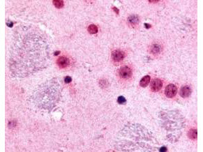
Rockland's Affinity Purified anti-mouse Gli-2 antibody was used at 10 μg/ml to evaluate staining on several mouse tissues. Moderate to strong staining was seen on many tissues with low background staining. This image shows Gli-2 staining of mouse brain. Tissue was formalin-fixed and paraffin embedded.
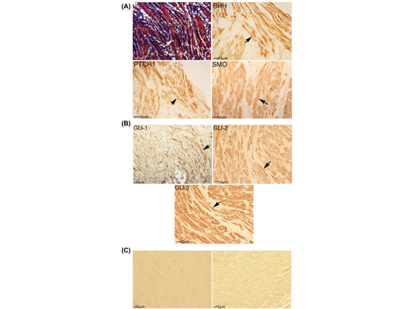
Trichrome stain and IHC analysis for SHH pathway in human RS muscle. A, Trichrome stain of human RS indicates abundant muscle and collagen. Arrows indicate muscle. IHC analysis of human RS shows SHH, PTCH1, and SMO protein localization in RS muscle. B, GLI-1, GLI-2 (p/n 600-401-695), and GLI-3 (p/n 600-401-694) are abundant in RS muscle. C, No primary controls (left: mouse anti-rabbit, and right: donkey anti-goat) are presented. Arrows indicate protein. 100?200× magnification. Fig 1. PMID:?30187971
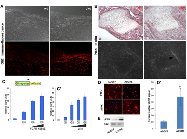
Effect of?Pten?deletion on hedgehog signaling.?(A) Immunofluorescence of GLI2 showed an increase in the expression of osteoblasts that line the perichondrium in the?Pten?conditional knockout (cko) compared with wild type (wt). (B) In situ hybridization for patched (Ptch1), a transcriptional target for GLI2, showed an increase (black arrow) in?Ptch1?expression in the cko compared with the wt control. (C,C′) Using a?Gli2?luciferase reporter plasmid transfected into C3H10T1/2 cells we observed that there is an activation of?Gli2?transcription when an active form of FGFR (FGFR-K650E) is co-transfected with GLI2. We also observed an increase in?Gli2?luciferase activity when we used an activated MEK kinase plasmid (repeated at least three times). Black wedges indicate increasing concentrations of FGFR-K650E or MEK co-transfected with Gli2. Luciferase activity was normalized to β-galactosidase activity. (D) Using?Pten?flox/flox calvarial osteoblasts, we used AdenoGFP (ADGFP) as control and AdenoCRE (ADCRE) to delete PTEN as can be observed in the indirect immunofluorescence images in the top panels. When PTEN is deleted we observe an increase in pERK nuclear localization. (D′) The number of cells with a nuclear pERK signal was determined revealing a significant increase in pERK (n=3, *P<0.005). Images are overlays of DAPI (blue) and rhodamine (red) staining. (E) Western blot data for pERK and total ERK from protein lysates of?Pten?flox/flox calvarial osteoblasts treated with AdenoCRE and AdenoGFP. All error bars indicate s.d. Fig 7. PMID:?21385768
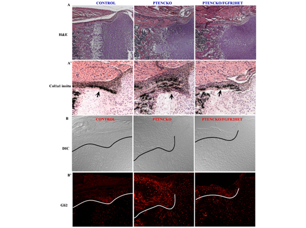
Rescue of the?Pten?conditional knockout (cko) mouse phenotype by deletion of one allele of?Fgfr2. (A,A′) Hematoxylin and Eosin (H&E) staining of the phenotype observed in the perichondrium, along with?Col1a1?in situ hybridization showing the increased perichondrium (black arrows) phenotype in the absence of?Pten?(PTENCKO). This phenotype is partially rescued with the deletion of?Pten?in the background of global loss of one allele of?Fgfr2?(PTENCKO/FGFR2HET). (B,B′) Immunohistochemistry for GLI2 protein levels in the perichondrium of tibial sections showed a similar increase in GLI2 protein levels in the absence of?Pten?but the GLI2 levels are comparable to the wild-type levels in the?Fgfr2?het?Pten?cko (PTENCKO/FGFR2HET) perichondrium. The conditional knockouts were generated using the?Dermo1cre mouse strain. DIC, differential interference contrast. Fig 8. PMID:?21385768
|
|
|
|
Rockland's Affinity Purified anti-mouse Gli-2 antibody was used at 10 μg/ml to evaluate staining on several mouse tissues. Moderate to strong staining was seen on many tissues, with low background staining. This image shows Gli-2 staining of mouse testis. Tissue was formalin-fixed and paraffin embedded.
|
|
| 別品名 |
rabbit anti-Gli-2 antibody, Gli 2, Gli2, zinc finger protein GLI2, Tax helper protein antibody, Thp antibody
|
| 交差種 |
Mouse
|
| 適用 |
Western Blot
Enzyme Linked Immunosorbent Assay
Immunohistochemistry
|
| 免疫動物 |
Rabbit
|
| 標識物 |
Unlabeled
|
| 精製度 |
Affinity Purified
|
| GENE ID |
14633
|
| Accession No.(Gene/Protein) |
124487481, Q8K0K3
|
| Gene Symbol |
Gli2
|
| 参考文献 |
[[Cosmo Reference Collection]][Pub Med ID]21385768,30187971
|
| [注意事項] |
濃度はロットによって異なる可能性があります。メーカーDS及びCoAからご確認ください。
|
|
| メーカー |
品番 |
包装 |
|
RKL
|
600-401-695
|
100 UG
|
※表示価格について
| 当社在庫 |
なし
|
| 納期目安 |
約10日
|
| 保存温度 |
-20℃
|
|
※当社では商品情報の適切な管理に努めておりますが、表示される法規制情報は最新でない可能性があります。
また法規制情報の表示が無いものは、必ずしも法規制に非該当であることを示すものではありません。
商品のお届け前に最新の製品法規制情報をお求めの際はこちらへお問い合わせください。
|
※当社取り扱いの試薬・機器製品および受託サービス・創薬支援サービス(納品物、解析データ等)は、研究用としてのみ販売しております。
人や動物の医療用・臨床診断用・食品用としては、使用しないように、十分ご注意ください。
法規制欄に体外診断用医薬品と記載のものは除きます。
|
|
※リンク先での文献等のダウンロードに際しましては、掲載元の規約遵守をお願いします。
|
|
※CAS Registry Numbers have not been verified by CAS and may be inaccurate.
|


