|
※サムネイル画像をクリックすると拡大画像が表示されます。
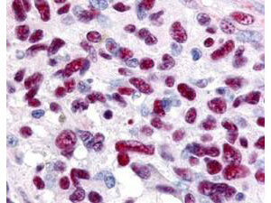
Immunohistochemistry of Rabbit anti-Gli-3 antibody. This image tissue: human glioblastoma. Specific staining was also noted in tissue from adrenal, brain, glioblastoma, colon, heart, kidney, lung, liver, skeletal muscle, ovary, pancreas, placenta, skin, spleen, stomach, testes, thymus, thyroid, tonsil and uterus. Fixation: formalin fixed paraffin embedded. Antigen retrieval: not required. Primary antibody: Gli-3 antibody at 0.625 μg/ml for 1 h at RT. Secondary antibody: Peroxidase rabbit secondary antibody at 1:10,000 for 45 min at RT. Localization: Gli-3 is nuclear and smooth muscle. Staining: Gli-3 as precipitated red signal with hematoxylin purple nuclear counterstain.
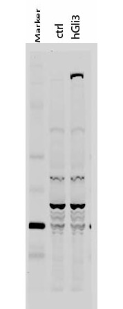
Western Blot of Rabbit anti-Gli-3 antibody. Lane 1: 50 kDa molecular weight marker. Lane 2: 293T cells transfected with CrkL-Flag. Lane 3: 293T cells transfected with human Gli-3. Load: 35 μg per lane. Primary antibody: Gli-3 antibody at 1:400 for overnight at 4°C. Secondary antibody: IRDye800TM rabbit secondary antibody at 1:10,000 for 45 min at RT. Block: 5% BLOTTO overnight at 4°C. Predicted/Observed size: 170-190 kDa for hGli-3. Other band(s): Non specific background ~60kDa.
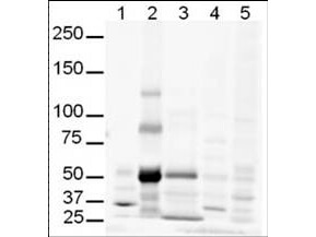
Western Blot of Rabbit anti-Gli-3 antibody. Lane 1: human brain whole cell lysate. Lane 2: human lung whole cell lysate. Lane 3: human spleen whole cell lysate. Lane 4: mouse brain whole cell lysate (p/n W10-000-T004). Lane 5: mouse lung whole cell lysate (p/n W10-000-MQ1). Load: 20 μg per lane. Primary antibody: Gli-3 antibody at 1:500 for overnight at 4°C. Secondary antibody: IRDye800TM rabbit secondary antibody at 1:10,000 for 45 min at RT. Block: 5% BLOTTO overnight at 4°C. Predicted/Observed size: Isoforms at ~170-190kDa and ~80kDa. Lane 2 shows what may be truncated Gli-3 (~80kDa). Other band(s): The strong band at ~50 kDa is unknown.
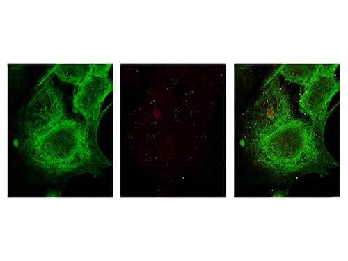
Immunofluorescence Microscopy of Rabbit anti-Gli-3 antibody. Tissue: MCF-7 cell. Antigen retrieval: not required. Primary antibody: Gli-3 antibody and Anti-alpha-Tubulin at 5 μg/mL for 1 h at RT. Secondary antibody: Fluorescein secondary antibody at 1:10,000 for 45 min at RT. Localization: Gli-3 is nuclear. Staining: Gli-3 staining as red fluorescent signal and Anti-alpha-Tubulin staining as green fluorescent signal using STED.
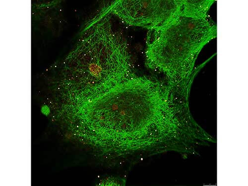
Immunofluorescence Microscopy of Rabbit anti-Gli-3 antibody. Tissue: MCF-7 cell. Antigen retrieval: not required. Primary antibody: Gli-3 antibody and Anti-alpha-Tubulin at 5 μg/mL for 1 h at RT. Secondary antibody: Fluorescein secondary antibody at 1:10,000 for 45 min at RT. Localization: Gli-3 is nuclear. Staining: Image (1) shows alpha-Tubulin staining as green fluorescent signal. Image (2) shows Gli-3 staining as red fluorescent signal and Images (3) shows both antibodies fluorescing using STED microscopy.
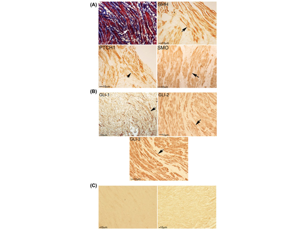
Trichrome stain and IHC analysis for SHH pathway in human RS muscle. A, Trichrome stain of human RS indicates abundant muscle and collagen. Arrows indicate muscle. IHC analysis of human RS shows SHH, PTCH1, and SMO protein localization in RS muscle. B, GLI-1, GLI-2 (p/n 600-401-695), and GLI-3 (p/n 600-401-694) are abundant in RS muscle. C, No primary controls (left: mouse anti-rabbit, and right: donkey anti-goat) are presented. Arrows indicate protein. 100?200× magnification. Fig 1. PMID:?30187971
|
|
|
|
Immunohistochemistry of Rabbit anti-Gli-3 antibody. This image tissue: human glioblastoma. Specific staining was also noted in tissue from adrenal, brain, glioblastoma, colon, heart, kidney, lung, liver, skeletal muscle, ovary, pancreas, placenta, skin, spleen, stomach, testes, thymus, thyroid, tonsil and uterus. Fixation: formalin fixed paraffin embedded. Antigen retrieval: not required. Primary antibody: Gli-3 antibody at 0.625 μg/ml for 1 h at RT. Secondary antibody: Peroxidase rabbit secondary antibody at 1:10,000 for 45 min at RT. Localization: Gli-3 is nuclear and smooth muscle. Staining: Gli-3 as precipitated red signal with hematoxylin purple nuclear counterstain.
|
|
| 別品名 |
Rabbit anti-GLI-3 antibody, Transcriptional activator GLI3, Gli 3, GLI3 form of 190 kDa, GLI3 form of 83 kDa
|
| 交差種 |
Human
|
| 適用 |
Western Blot
Enzyme Linked Immunosorbent Assay
Immunohistochemistry
Immuno Fluorescence
|
| 免疫動物 |
Rabbit
|
| 抗原部位 |
a.a.41-57
|
| 標識物 |
Unlabeled
|
| 精製度 |
Affinity Purified
|
| GENE ID |
2737
|
| Accession No.(Gene/Protein) |
119393899, P10071
|
| Gene Symbol |
GLI3
|
| 参考文献 |
[[Cosmo Reference Collection]][Pub Med ID]30187971
|
| [注意事項] |
濃度はロットによって異なる可能性があります。メーカーDS及びCoAからご確認ください。
|
|
| メーカー |
品番 |
包装 |
|
RKL
|
600-401-694
|
100 UG
|
※表示価格について
| 当社在庫 |
なし
|
| 納期目安 |
約10日
|
| 保存温度 |
-20℃
|
|


