|
※サムネイル画像をクリックすると拡大画像が表示されます。
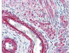
Rockland's affinity purified anti-Monophosphorylated RLC Smooth and Non-Muscle Myosin pS19/20 antibody was used at 2.5 μg/ml to detect signal in a variety of tissues including multi-human, multi-brain and multi-cancer slides. This image shows strong staining of both vascular and myometrial smooth muscle cells of the uterus. Tissue was formalin-fixed and paraffin embedded. The image shows localization of the antibody as the precipitated red signal, with a hematoxylin purple nuclear counterstain. Personal Communication, Tina Roush, LifeSpanBiosciences, Seattle, WA.
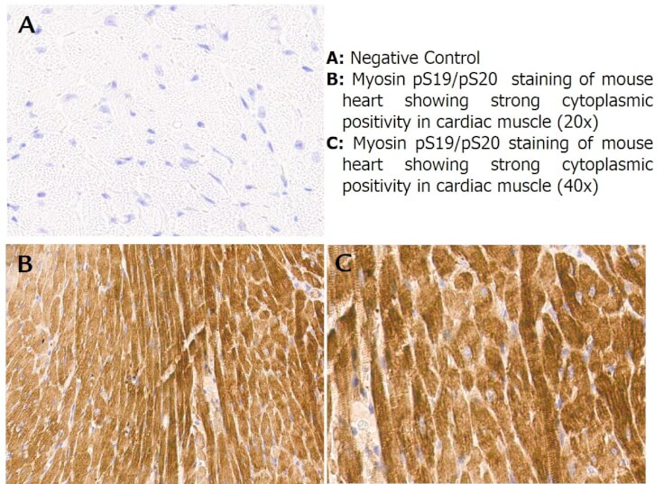
Immunohistochemistry with anti-myosin pS19/pS20 antibody showing strong cytoplasmic staining of myocytes in mouse heart muscle 20x and 40x (B & C). Staining was performed on Leica Bond system using the standard protocol. Formalin fixed/paraffin embedded tissue sections were subjected to antigen retrieval and then incubated with rabbit anti-myosin pS19/pS20 antibody at 1:100 dilution for 60 minutes. Biotinylated Anti-rabbit secondary antibody was used to detect primary antibody. The reaction was developed using streptavidin-HRP conjugated compact polymer system and visualized with chromogen substrate, 3’3-diamino-benzidine substrate (DAB). The sections were then counterstained with hematoxylin to detect cell nuclei.
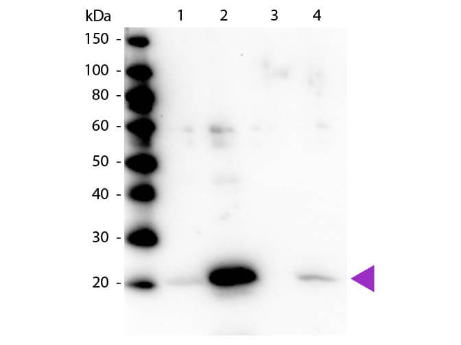
Western blot of Rabbit Anti-Myosin pS19/pS20 primary antibody. Lane 1: Regulatory Light Chain Non-Phospho recombinant protein. Lane 2: Regulatory Light Chain Phospho recombinant protein. Lane 3: Smooth Muscle Non-Phospho recombinant protein. Lane 4: Smooth Muscle Phospho recombinant protein. Load: 50 ng per lane. Primary antibody: Myosin pS19/pS20 primary antibody at 1:1,000 overnight at 4?C. Secondary antibody: Peroxidase rabbit secondary antibody at 1:40,000 for 60 min at RT. Blocking: MB-070 for 30 min at RT. Predicted/Observed size: 20 kDa, 20 kDa for Regulatory Light Chain Phospho. Other band(s): None.
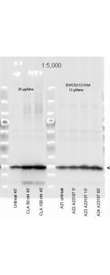
Affinity Purified Phospho specific antibody to Monophosphorylated Regulatory Light Chain of Smooth and Non-muscle Myosin at pS19/pS20 was used at a 1:5000 dilution to detect myosin light chain by Western blot. Either 13μL or 20 μg of a mouse cardiac myocyte lysate was loaded on a 4-20% Criterion gel for SDS-PAGE. Samples were either mock-treated or CLA-treated, as indicated. After washing, a 1:5,000 dilution of HRP conjugated Gt-a-Rabbit IgG (611-103-122) preceded color development using Amersham's substrate system. Other detection methods will yield similar results. Data courtesy of the Alliance for Cellular Signaling (http://www.signaling-gateway.org).
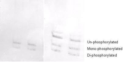
Affinity purified phosphospecific antibody to phosphorylated regulatory light chain of smooth and non-muscle Myosin at pS19/pS20 was used at a 1:1000 dilution to detect myosin light chain by Western blot on 3T3 cell lysates. A standard urea/glycerol gel without SDS was used to separate phospho forms of regulatory light chain according to mass to charge ratios. In Panel A on the left, reactivity of Rockland's phosphospecific antibody is shown. In Panel B on the right, reactivity of commercially available pan reactive antibody that detects both un-phosphorylated and phosphorylated forms of regulatory light chain is shown. Rockland's phosphospecific antibody detects both mono-phosphorylated (pSer20 Mono-P-RLC) and di-phosphorylated (pThr19-pSer20 Di-P-RLC) regulatory light chain. Personal communication. J. Stull. UT Southwestern Medical Center.
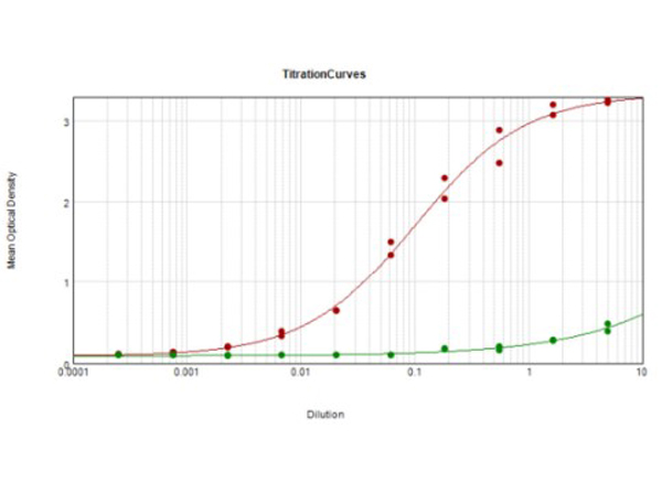
ELISA Results of Rabbit Anti-Myosin pS19/pS20 Antibody tested against BSA-conjugated peptide of immunizing peptide. Each well was coated in duplicate with 0.1μg of Myosin pS19/pS20 [Red Line] and Myosin S19/S20 [Green Line]. The starting dilution of antibody was 5μg/ml and the X-axis represents the Log10 of a 3-fold dilution. This titration is a 4-parameter curve fit where the IC50 is defined as the titer of the antibody. Assay performed using Goat anti-Rabbit IgG Antibody Peroxidase Conjugated (Min X Bv Ch Gt GP Ham Hs Hu Ms Rt & Sh Serum Proteins) (p/n 611-103-122) and TMB ELISA Peroxidase Substrate (p/n TMBE-1000).
|
|
|
|
Rockland's affinity purified anti-Monophosphorylated RLC Smooth and Non-Muscle Myosin pS19/20 antibody was used at 2.5 μg/ml to detect signal in a variety of tissues including multi-human, multi-brain and multi-cancer slides. This image shows strong staining of both vascular and myometrial smooth muscle cells of the uterus. Tissue was formalin-fixed and paraffin embedded. The image shows localization of the antibody as the precipitated red signal, with a hematoxylin purple nuclear counterstain. Personal Communication, Tina Roush, LifeSpanBiosciences, Seattle, WA.
|
|
| 別品名 |
rabbit anti-Myosin p19/pS20 antibody, Myosin regulatory light chain 12A, Myosin regulatory light chain MRLC3, Myosin regulatory light chain 2 nonsarcomeric, Myosin RLC, MLC-2B, HEL-S-24, Epididymis secretory protein Li 24, MLCB
|
| 交差種 |
Human
Mouse
|
| 適用 |
Western Blot
Enzyme Linked Immunosorbent Assay
Immunohistochemistry
|
| 免疫動物 |
Rabbit
|
| 抗原部位 |
N-terminus
|
| 標識物 |
Unlabeled
|
| 精製度 |
Affinity Purified
|
| 翻訳後修飾 |
リン酸化
|
| GENE ID |
10627
|
| Accession No.(Gene/Protein) |
AAH16372.1, P19105
|
| Gene Symbol |
MYL12A
|
| 参考文献 |
[Pub Med ID]25637353
|
| [注意事項] |
濃度はロットによって異なる可能性があります。メーカーDS及びCoAからご確認ください。
|
|
| メーカー |
品番 |
包装 |
|
RKL
|
600-401-416
|
100 UG
|
※表示価格について
| 当社在庫 |
なし
|
| 納期目安 |
約10日
|
| 保存温度 |
-20℃
|
|
※当社では商品情報の適切な管理に努めておりますが、表示される法規制情報は最新でない可能性があります。
また法規制情報の表示が無いものは、必ずしも法規制に非該当であることを示すものではありません。
商品のお届け前に最新の製品法規制情報をお求めの際はこちらへお問い合わせください。
|
※当社取り扱いの試薬・機器製品および受託サービス・創薬支援サービス(納品物、解析データ等)は、研究用としてのみ販売しております。
人や動物の医療用・臨床診断用・食品用としては、使用しないように、十分ご注意ください。
法規制欄に体外診断用医薬品と記載のものは除きます。
|
|
※リンク先での文献等のダウンロードに際しましては、掲載元の規約遵守をお願いします。
|
|
※CAS Registry Numbers have not been verified by CAS and may be inaccurate.
|
