

| 別品名 |
Ataxia telangiectasia mutated
|
| 抗原部位 |
a.a.1974-1988
|
| 種由来 |
Human
|
| 標識物 |
Unlabeled
|
| 精製度 |
Affinity Purified
|
| 適用 |
Western Blot
Enzyme Linked Immunosorbent Assay
Immunohistochemistry
|
| 免疫動物 |
Rabbit
|
| 交差種 |
Human
Mouse
|
| GENE ID |
472
|
| Accession No.(Gene/Protein) |
NP_000042, Q13315
|
| Gene Symbol |
ATM
|
| 形状 |
滅菌済み液状品
|
| 参考文献 |
[Pub Med ID]15964794, 22826323, 21464229, 17126333, 22797809, 26649750, 21554499
|
| [注意事項] |
濃度はロットによって異なる可能性があります。メーカーDS及びCoAからご確認ください。
|
|
※サムネイル画像をクリックすると拡大画像が表示されます。
DNA DSBs induced by Q6 triggered ATM Chk2 pathway in hypoxia.Western blot was carried out to explore the ATM/ATR signaling pathways in respond to DNA DSBs induced by Q6. A. HepG2 cells and B. Bel 7402 cells were treated with different concentration of Q6 (2.5 μM, 5 μM) under hypoxia (1% O2) condition. Protein levels were detected by Western blot analysis. B Actin was measured as the loading control. Data are representative of three independent experiments. Figure provided by CiteAb. Source: PLoS One, PMID: 26649750.
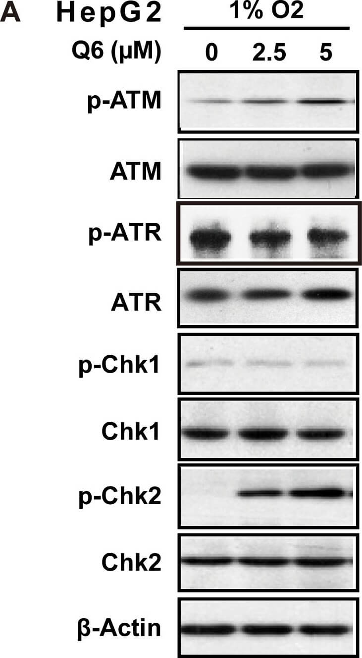
DNA DSBs induced by Q6 triggered ATM-Chk2 pathway in hypoxia.Western blot was carried out to explore the ATM/ATR signaling pathways in respond to DNA DSBs induced by Q6. A. HepG2 cells and B. Bel-7402 cells were treated with different concentration of Q6 (2.5 μM, 5 μM) under hypoxia (1% O2) condition. Protein levels were detected by Western blot analysis. β-Actin was measured as the loading control. Data are representative of three independent experiments. Figure provided by CiteAb. Source: PLoS One, PMID: 26649750.
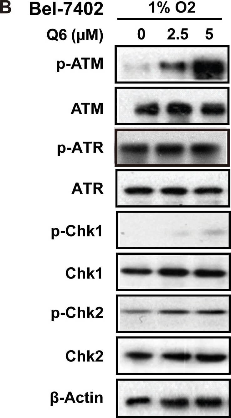
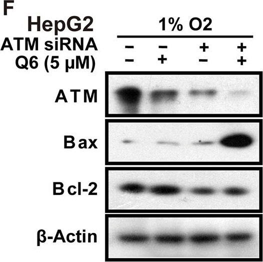
Q6 induced G2/M arrest and apoptosis is ATM/Chk2 dependent in hypoxia.A. HepG2 and Bel-7402 cells, treated with Q6 (5 μM) in the presence or absence of caffeine (2 mM) for 24 h under hypoxia (1% O2), were collected and prepared for cytometric analysis of cell cycle distribution. B & C. HepG2 cells treated with Q6 (10 μM) in the presence or absence of caffeine (2 mM) for 24 h under hypoxia (1% O2). Detection of apoptosis by flow cytometry (B) and caspase cascade by Western blot (C) were then performed. D & E. HepG2 cells treated with Q6 in the presence or absence of ATM specific inhibitor KU-60019 (3 μM) under hypoxia (1% O2), and subjected to sub-G1 analyses (D) and Western blot analyses (E), respectively. F. Western blot was used to assess the role of ATM during apoptosis induced by Q6 in hypoxia. HepG2 cells were treated with ATM RNAi or vector RNAi in the presence or absence of Q6 (5 μM) under hypoxia (1% O2) condition. Figure provided by CiteAb. Source: PLoS One, PMID: 26649750.
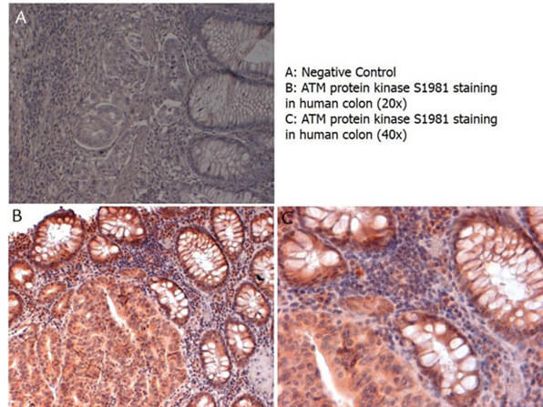
Immunohistochemistry with anti-ATM protein kinase S1981 antibody showing ATM protein kinase S1981 staining in nucleus and cytoplasm of human colon at 20x and 40x (B & C). Formalin fixed/paraffin embedded sections were subjected to heat induced epitope retrieval (HIER) at pH 6.2 and then incubated with rabbit anti-ATM protein kinase S1981 antibody at 4.0 ug/ml for 60 minutes. The reaction was developed using MACH 1 universal HRP polymer detection system and visualized with 3’3-diamino-benzidine substrate (DAB).
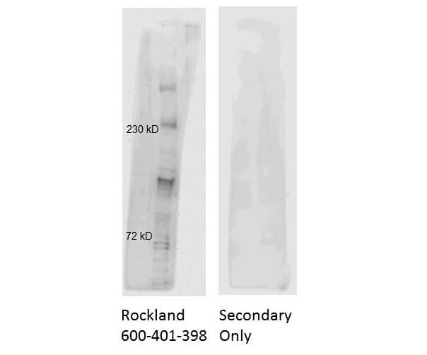
Western Blot of Rabbit Anti-ATM Antibody. HeLa Nuclear Lysates run on 4-8% gel and transferred for 1 hr at 100V to 0.45 um nitrocellulose. Antibody Dilution Buffer: Buffer (p/n MB-070). Block: 5% Blotto for 1hr at 2-8C. Primary Antibody: Anti-ATM at 1:1000 overnight at 2-8C. Secondary Antibody: Goat Anti-Rabbit IgG HRP (p/n 611-103-122) at 1:20,000 for 1hr at 2-8C.
|

|
|
DNA DSBs induced by Q6 triggered ATM Chk2 pathway in hypoxia.Western blot was carried out to explore the ATM/ATR signaling pathways in respond to DNA DSBs induced by Q6. A. HepG2 cells and B. Bel 7402 cells were treated with different concentration of Q6 (2.5 μM, 5 μM) under hypoxia (1% O2) condition. Protein levels were detected by Western blot analysis. B Actin was measured as the loading control. Data are representative of three independent experiments. Figure provided by CiteAb. Source: PLoS One, PMID: 26649750.
|
|
|
| メーカー |
品番 |
包装 |
|
RKL
|
600-401-398
|
100 UG
|
※表示価格について
| 当社在庫 |
なし
|
| 納期目安 |
約10日
|
| 保存温度 |
-20℃
|
|


