|
※サムネイル画像をクリックすると拡大画像が表示されます。
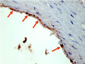
Rockland's Affinity Purified anti-HA epitope tag polyclonal antibody detects HA tagged recombinant proteins by IHC on formalin fixed paraffin embedded tissue. Arrowheads point to expression of HA tagged proteins in endothelial cells of mouse aorta. Sections of 4 μm were prepared from representative paraffin blocks. Sections were then deparaffinized and rehydrated with xylene and alcohol. Citrate buffer antigen retrieval was performed for 30 min in a boiling jar. Anti-HA was diluted in blocking buffer at 1:2,000 and reacted at 4° C overnight followed by signal detection using horseradish peroxidase with DAB as the chromogenic substrate. Tissue was counterstained with Mayer's hematoxylin. Personal Communication, Behzad Yeganeh,U. Manitoba, Winnipeg, Canada.
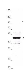
Anti-HA epitope tag polyclonal antibody detects HA-tagged recombinant proteins by western blot.? Polyclonal Rabbit anti-HA epitope tag, at a 1:2,000 dilution, was used to detect 1.0 μg of 12-Epitope Tag Protein Marker Lysate (p/n MB-301-0100) containing the HA epitope tag.?? A 4-20% gradient gel was used to resolve the protein by SDS-PAGE.? The lysate was transferred to nitrocellulose using standard methods.? After blocking, the membrane was probed with Rockland's anti-HA tag antibody for 1 h at room temperature followed by washes and reaction with a 1:20,000 dilution of IRDyeR 800 conjugated Gt-a-Rabbit IgG (H&L) MX10 (code 611-132-122) for 30 min at room temperature.? LICOR's OdysseyR Infrared Imaging System was used to scan and process the image.? Other detection systems will yield similar results.
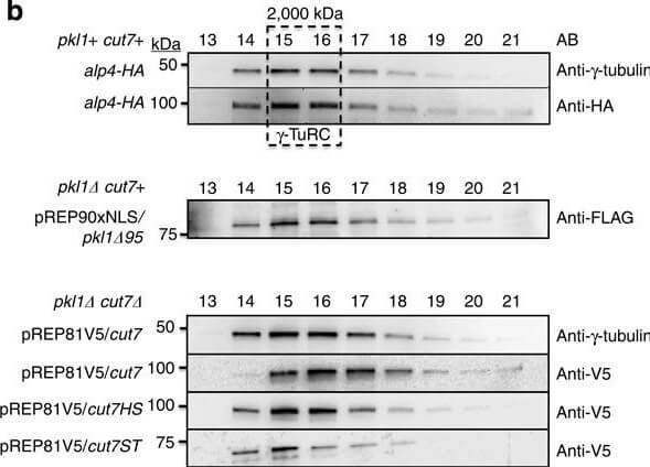
Kinesin-5 Cut7 binds the γ-TuRC MTOC.(a) Kinesin-5 and kinesin-14 constructs used in Fast Protein Liquid Chromatography. V5-tagged Cut7 and two truncation constructs were used, in addition to one FLAG-Pkl1 truncated construct that retains full Pkl1 activity. Cut7 constructs are V5-tagged full-length Cut7 (aa 1?1,085), Cut7-Head-Stalk (Cut7HS, aa 1?888) and Cut7-Stalk-Tail (Cut7-ST, aa 443?1,085). (b) Western blot profiles of whole-cell extracts fractionated by Separose 6 using FPLC. (c) Western blots of Cut7 constructs immunoprecipitated from whole-cell extracts using anti-V5 magnetic beads with empty strain negative controls. (d) Cartoon diagram of 6-His tagged Pkl1 Tail peptide co-immunoprecipitation assay using magnetic beads with His affinity and FPLC fraction 15. (e) Pkl1 Tail peptide co-immunoprecipitation of γ-TuRC core subunits and V5-Cut7ST using a short Pkl1 Tail peptide (PγT). Mutated peptide PγM has significantly reduced interaction with the fission yeast γ-TuRC. The anti-HA antibody detects the HA-tagged γ-TuRC protein Alp4. Figure provided by CiteAb. Source: Nat Commun, PMID: 25348260.
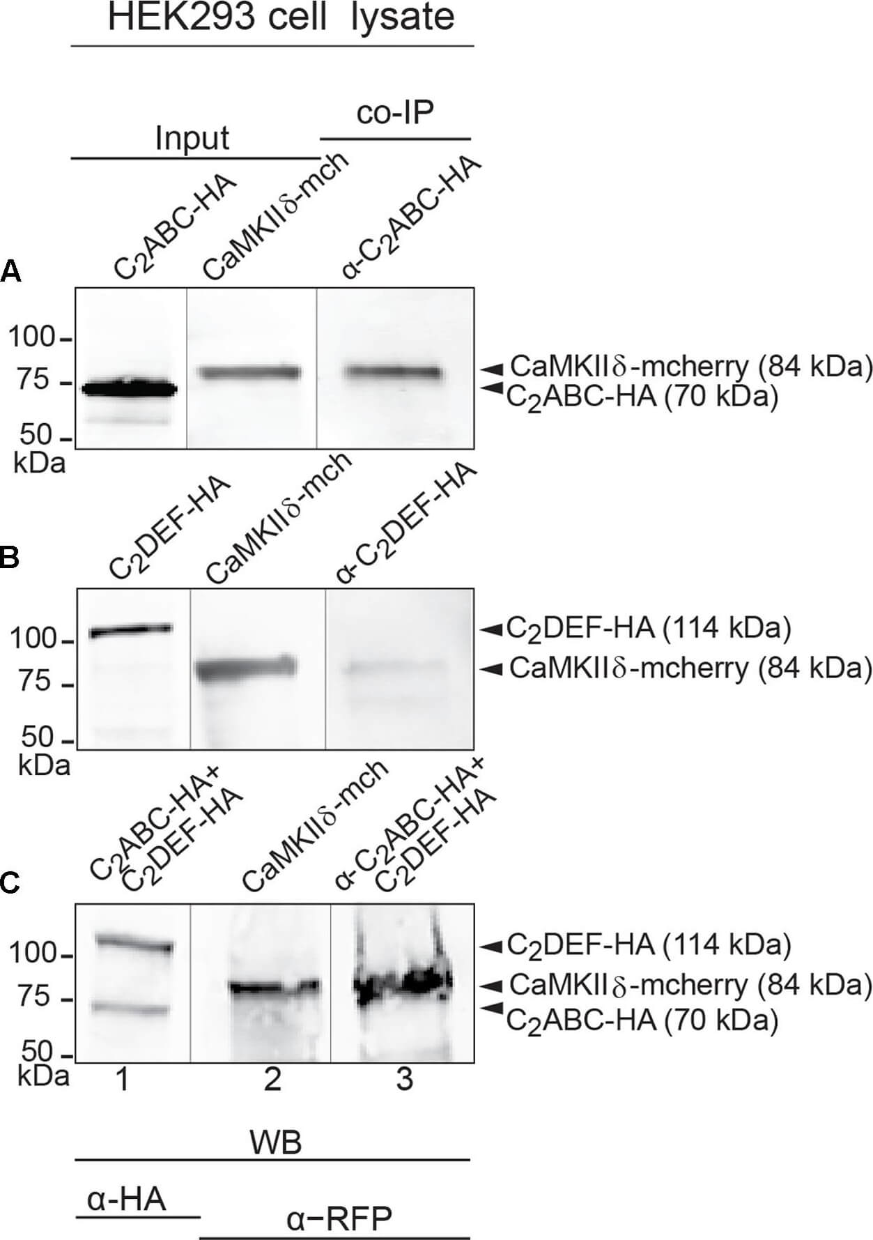
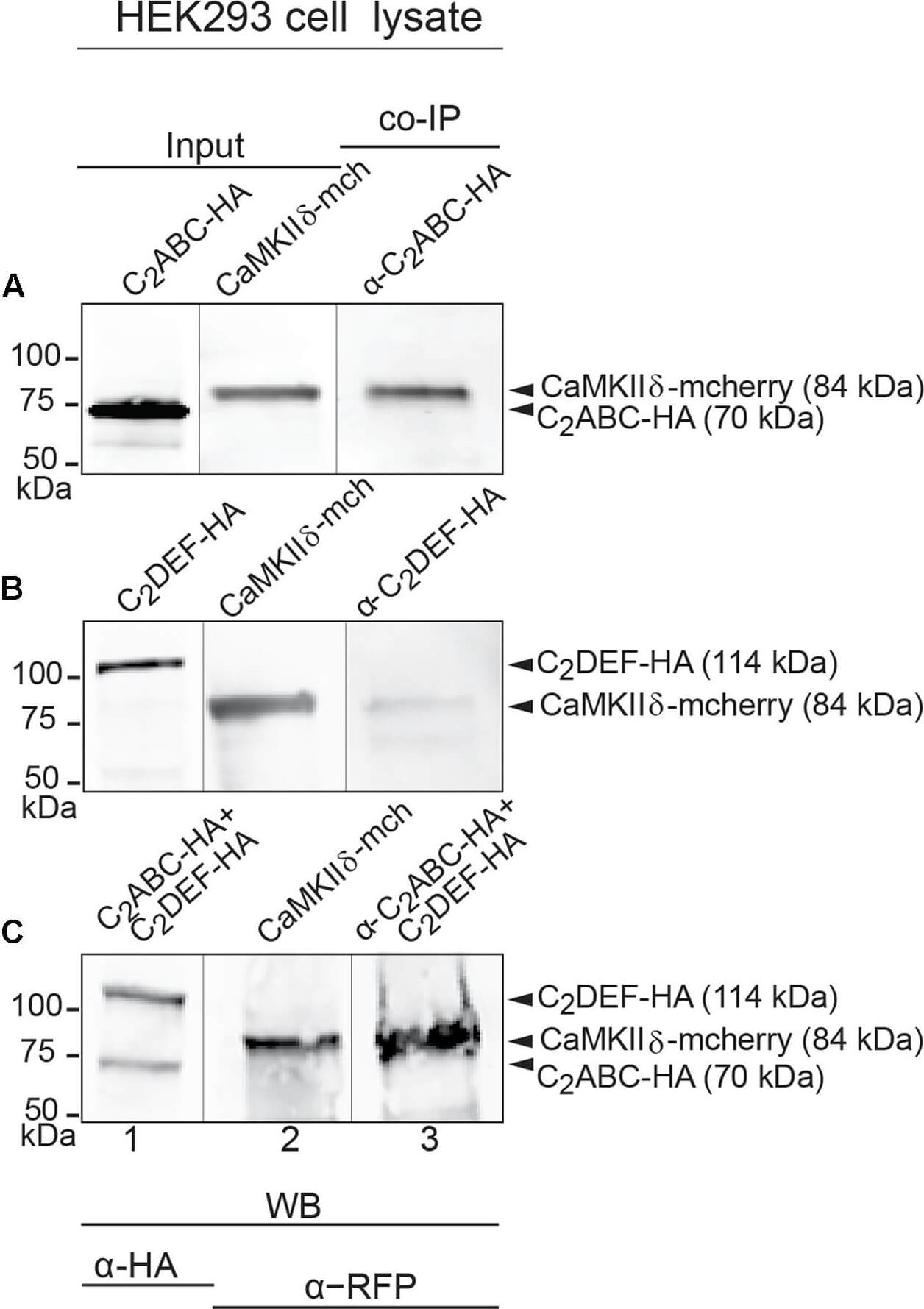
Immunoprecipitation and western blot show interaction of otoferlin with CaMKIIδ. (A?C) Two HA-tagged mouse otoferlin fragments, C2ABC (aa 1?632 in NP_001093865; 70 kDa) and C2DEF (aa 933?1920; 114 kDa) were co-transfected with mcherry-tagged mouse CaMKIIδ into HEK293 cells. Transfections were performed either with otoferlin C2ABC and CaMKIIδ (A, Input Lane 1 and 2), otoferlin C2DEF and CaMKIIδ (B, Input Lane 1 and 2) or in the presence of both C2ABC and C2DEF fragments and CaMKIIδ (C, Input Lane 1 and 2). Co-immunoprecipitations of C2ABC-HA and C2DEF-HA were conducted from HEK293 cell lysates using anti-HA antibodies. CaMKIIδ-mcherry was detected in the eluate using an anti-RFP (red fluorescent protein) antibody (A?C, Lane 3), indicating that CaMKIIδ co-precipitated with recombinant otoferlin fragments. Figure provided by CiteAb. Source: Front Synaptic Neurosci, PMID: 29046633.
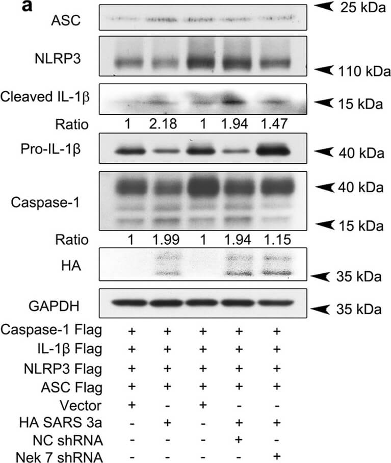
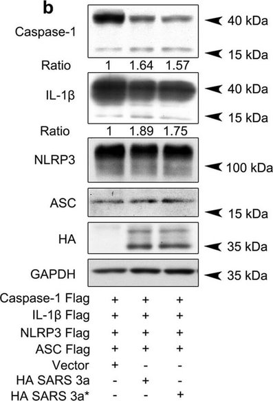
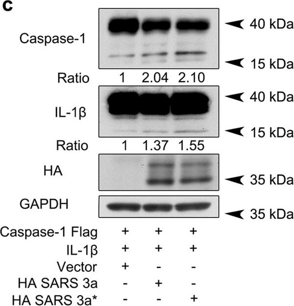
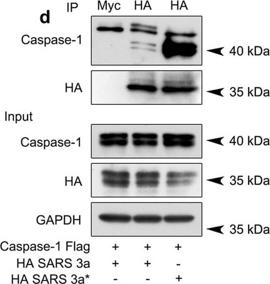
SARS 3a induces NLRP3 inflammasome activation by multiple mechanisms. A) Immunoblot analysis of the pro- and cleaved forms of caspase-1 and IL-1β after reconstitution of inflammasome in HEK 293T cells transfected with SARS 3a with or without NEK7 shRNA. B) Immunoblot analysis of the pro- and cleaved forms of caspase-1 and IL-1β after reconstitution of inflammasome and transfection with SARS 3a or SARS 3a C133A. C) Immunoblot analysis of the pro- and cleaved forms of caspase-1 and IL-1β after co-transfection with caspase-1, IL-1β, and SARS 3a or SARS 3a C133A. d Immunoprecipitation analysis of interaction between SARS 3a or SARS 3a C133A and caspase-1. All western blot data are representative of two or three independent experiments Figure provided by CiteAb. Source: Cell Death Dis, PMID: 30185776.
SARS 3a induces NLRP3 inflammasome activation by multiple mechanisms. A) Immunoblot analysis of the pro- and cleaved forms of caspase-1 and IL-1β after reconstitution of inflammasome in HEK 293T cells transfected with SARS 3a with or without NEK7 shRNA. B) Immunoblot analysis of the pro- and cleaved forms of caspase-1 and IL-1β after reconstitution of inflammasome and transfection with SARS 3a or SARS 3a C133A. C) Immunoblot analysis of the pro- and cleaved forms of caspase-1 and IL-1β after co-transfection with caspase-1, IL-1β, and SARS 3a or SARS 3a C133A. D) Immunoprecipitation analysis of interaction between SARS 3a or SARS 3a C133A and caspase-1. All western blot data are representative of two or three independent experiments Figure provided by CiteAb. Source: Cell Death Dis, PMID: 30185776.
SARS 3a induces NLRP3 inflammasome activation by multiple mechanisms. A) Immunoblot analysis of the pro- and cleaved forms of caspase-1 and IL-1β after reconstitution of inflammasome in HEK 293T cells transfected with SARS 3a with or without NEK7 shRNA. B) Immunoblot analysis of the pro- and cleaved forms of caspase-1 and IL-1β after reconstitution of inflammasome and transfection with SARS 3a or SARS 3a C133A. C) Immunoblot analysis of the pro- and cleaved forms of caspase-1 and IL-1β after co-transfection with caspase-1, IL-1β, and SARS 3a or SARS 3a C133A. D) Immunoprecipitation analysis of interaction between SARS 3a or SARS 3a C133A and caspase-1. All western blot data are representative of two or three independent experiments Figure provided by CiteAb. Source: Cell Death Dis, PMID: 30185776.
SARS 3a induces NLRP3 inflammasome activation by multiple mechanisms. A) Immunoblot analysis of the pro- and cleaved forms of caspase-1 and IL-1β after reconstitution of inflammasome in HEK 293T cells transfected with SARS 3a with or without NEK7 shRNA. B) Immunoblot analysis of the pro- and cleaved forms of caspase-1 and IL-1β after reconstitution of inflammasome and transfection with SARS 3a or SARS 3a C133A. C) Immunoblot analysis of the pro- and cleaved forms of caspase-1 and IL-1β after co-transfection with caspase-1, IL-1β, and SARS 3a or SARS 3a C133A. D) Immunoprecipitation analysis of interaction between SARS 3a or SARS 3a C133A and caspase-1. All western blot data are representative of two or three independent experiments Figure provided by CiteAb. Source: Cell Death Dis, PMID: 30185776.
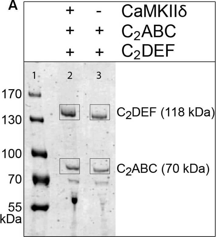
Otoferlin is phosphorylated by CaMKIIδ in vitro. (A) Otoferlin fragments C2ABC (aa 1?616 in NP_001093865, 70 kDa) and C2DEF (aa 908?1932, 118 kDa), were expressed in E. coli and subjected to an in vitro phosphorylation assay with CaMKIIδ and Ca2+/calmodulin. Reactions were stopped after 5 min of incubation and proteins were run on a Coomassie gel. Note the slight shift in mass of the fragments between experiment (lane 2) and control without kinase (lane 3). Coomassie stained bands corresponding to otoferlin C2DEF and C2ABC were cut off the gel and processed for mass spectrometric analysis of otoferlin phosphorylation (Supplementary Figure S2). (B) Three independent experiments as in (A) revealed 10 serine/threonines in otoferlin that were reproducibly phosphorylated by CaMKIIδ. The putative otoferlin domain topology (in mouse isoform 1; NP_001093865) predicts six C2 domains (C2A to C2F; purple), a coiled-coiled domain (orange), a FerB domain (yellow), and a transmembrane domain (TM) (dark gray). Five of the phosphorylation sites are located in C2 domains. Figure provided by CiteAb. Source: Front Synaptic Neurosci, PMID: 29046633.
Immunoprecipitation and western blot show interaction of otoferlin with CaMKIIδ. (A?C) Two HA-tagged mouse otoferlin fragments, C2ABC (aa 1?632 in NP_001093865; 70 kDa) and C2DEF (aa 933?1920; 114 kDa) were co-transfected with mcherry-tagged mouse CaMKIIδ into HEK293 cells. Transfections were performed either with otoferlin C2ABC and CaMKIIδ (A, Input Lane 1 and 2), otoferlin C2DEF and CaMKIIδ (B, Input Lane 1 and 2) or in the presence of both C2ABC and C2DEF fragments and CaMKIIδ (C, Input Lane 1 and 2). Co-immunoprecipitations of C2ABC-HA and C2DEF-HA were conducted from HEK293 cell lysates using anti-HA antibodies. CaMKIIδ-mcherry was detected in the eluate using an anti-RFP (red fluorescent protein) antibody (A?C, Lane 3), indicating that CaMKIIδ co-precipitated with recombinant otoferlin fragments. Figure provided by CiteAb. Source: Front Synaptic Neurosci, PMID: 29046633.
|
|
|
|
Rockland's Affinity Purified anti-HA epitope tag polyclonal antibody detects HA tagged recombinant proteins by IHC on formalin fixed paraffin embedded tissue. Arrowheads point to expression of HA tagged proteins in endothelial cells of mouse aorta. Sections of 4 μm were prepared from representative paraffin blocks. Sections were then deparaffinized and rehydrated with xylene and alcohol. Citrate buffer antigen retrieval was performed for 30 min in a boiling jar. Anti-HA was diluted in blocking buffer at 1:2,000 and reacted at 4° C overnight followed by signal detection using horseradish peroxidase with DAB as the chromogenic substrate. Tissue was counterstained with Mayer's hematoxylin. Personal Communication, Behzad Yeganeh,U. Manitoba, Winnipeg, Canada.
|
|
| 別品名 |
rabbit anti-HA epitope tag antibody, rabbit anti-hemagglutinin antibody, rabbit anti-HA tag antibody, anti-epitope
|
| 適用 |
Western Blot
Enzyme Linked Immunosorbent Assay
Immunohistochemistry
|
| 免疫動物 |
Rabbit
|
| 抗原部位 |
a.a.114-122, YPYDVPDYA
|
| 標識物 |
Unlabeled
|
| 精製度 |
Affinity Purified
|
| Tag情報 |
HA
|
| 参考文献 |
[Pub Med ID]32156817
|
| [注意事項] |
濃度はロットによって異なる可能性があります。メーカーDS及びCoAからご確認ください。
|
|
| メーカー |
品番 |
包装 |
|
RKL
|
600-401-384
|
100 UG
|
※表示価格について
| 当社在庫 |
なし
|
| 納期目安 |
約10日
|
| 保存温度 |
-20℃
|
|
