|
※サムネイル画像をクリックすると拡大画像が表示されます。
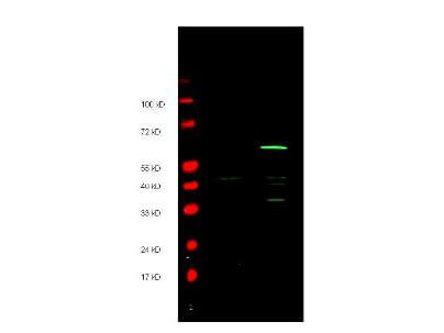
Anti-Myc epitope tag polyclonal antibody detects both AMINO and CARBOXY terminal linked Myc-tagged recombinant proteins by western blot. Polyclonal rabbit host anti-Myc epitope tag antibody was diluted to 1.0 μg/ml to detect either recombinant protein.? 4-20% gradient gels were used to resolve the proteins by SDS-PAGE.? The proteins were transferred to nitrocellulose using standard methods.? After blocking, the membranes were probed with the primary antibody overnight at 4°C followed by washes and reaction with a 1:10,000 dilution of IRDyeR 800 conjugated Gt-a-Rabbit IgG (H&L) MX10 (code 611-132-122) for 45 min at room temperature (Green, 800 nm channel).?? Pre-stained molecular weight markers are also shown (lane M, Red, 700 nm channel).? LICOR's OdysseyR Infrared Imaging System was used to scan and process the image.? Other detection systems will yield similar results.
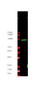
Anti-Myc epitope tag polyclonal antibody detects ~ 100 kDa CARBOXY terminal linked Myc-tagged recombinant protein present in ~35 μg of lysate by western blot. Carboxy terminal linked Myc recombinant protein was the gift of Zhongsheng You, Salk Institute, LaJolla, CA.
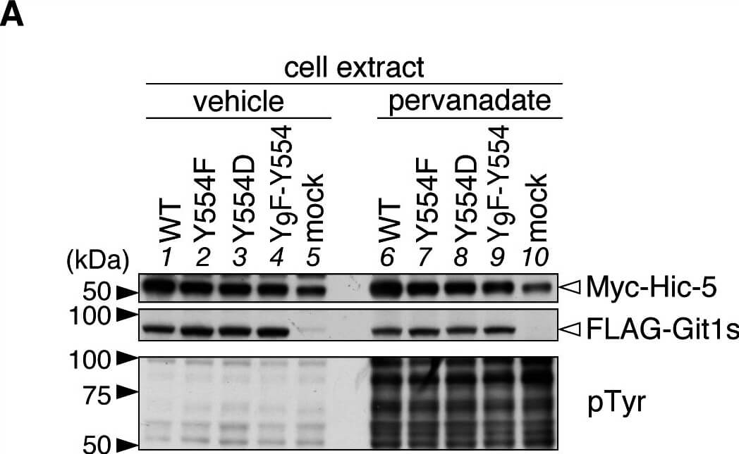
Git1 phosphorylation at Tyr-554 weakened its association with Hic-5.A, Western blotting of protein expression levels, and tyrosine phosphorylation of all proteins in HEK293T cells expressing FLAG-tagged Git1 proteins (Fig. 1A) together with Myc-tagged Hic-5. Cells were treated with 100 μM pervanadate or vehicle for 15 min, and then analyzed by Western blotting using anti-FLAG M2, anti-Myc 9E10, or anti-phosphotyrosine PY20. B, Co-immunoprecipitaion of Git1 mutants with Hic-5. The immunoprecipitates from cell extracts with anti-FLAG beads were analyzed by Western blotting with an anti-FLAG or anti-Myc antibody. To verify the tyrosine phosphorylation of FLAG-tagged Git1 proteins, the same membrane was reacted with anti-phosphotyrosine PY20. Ig, immunoglobulin. The lower part shows the densitometric analysis of the relative amount of Myc-Hic-5 to FLAG-Git1 in the immunoprecipitates. Data are the mean ± S.E. (error bars; n = 3). **, P < 0.01 significantly different from the wild-type with the same treatment; #, P < 0.05 or , P < 0.01 significant difference between vehicle- and pervanadate-treated groups by ANOVA with Fisher’s PLSD post hoc tests. Figure provided by CiteAb. Source: PLoS One, PMID: 25742295.
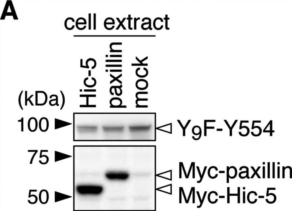
Git1 phosphorylation at Tyr-554 was enhanced by co-expression of paxillin.A, Western blotting of protein expression levels in HEK293T cells exogenously expressing the FLAG-tagged Git1-Y9F-Y554 mutant together with Myc-tagged paxillin, Myc-tagged Hic-5, or a control mock. B, Tyrosine phosphorylation of Y9F-Git1 proteins in anti-FLAG immunoprecipitates. The lower graph shows the densitometric analysis of the Western blotting data. Data are the mean ± S.E. (error bars; n = 3). *, P < 0.05 significantly different from Hic-5-transfected cells by ANOVA with Fisher’s PLSD post hoc tests. Figure provided by CiteAb. Source: PLoS One, PMID: 25742295.
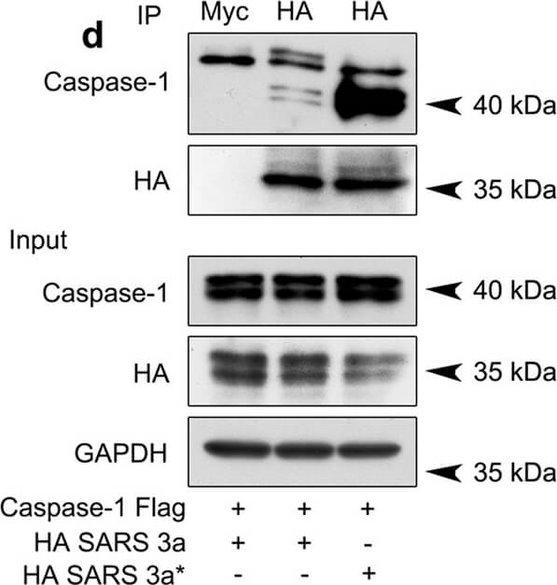
SARS 3a induces NLRP3 inflammasome activation by multiple mechanisms. A) Immunoblot analysis of the pro- and cleaved forms of caspase-1 and IL-1β after reconstitution of inflammasome in HEK 293T cells transfected with SARS 3a with or without NEK7 shRNA. B) Immunoblot analysis of the pro- and cleaved forms of caspase-1 and IL-1β after reconstitution of inflammasome and transfection with SARS 3a or SARS 3a C133A. C) Immunoblot analysis of the pro- and cleaved forms of caspase-1 and IL-1β after co-transfection with caspase-1, IL-1β, and SARS 3a or SARS 3a C133A. D) Immunoprecipitation analysis of interaction between SARS 3a or SARS 3a C133A and caspase-1. All western blot data are representative of two or three independent experiments Figure provided by CiteAb. Source: Cell Death Dis, PMID: 30185776.
|
|
|
|
Anti-Myc epitope tag polyclonal antibody detects both AMINO and CARBOXY terminal linked Myc-tagged recombinant proteins by western blot. Polyclonal rabbit host anti-Myc epitope tag antibody was diluted to 1.0 μg/ml to detect either recombinant protein.? 4-20% gradient gels were used to resolve the proteins by SDS-PAGE.? The proteins were transferred to nitrocellulose using standard methods.? After blocking, the membranes were probed with the primary antibody overnight at 4°C followed by washes and reaction with a 1:10,000 dilution of IRDyeR 800 conjugated Gt-a-Rabbit IgG (H&L) MX10 (code 611-132-122) for 45 min at room temperature (Green, 800 nm channel).?? Pre-stained molecular weight markers are also shown (lane M, Red, 700 nm channel).? LICOR's OdysseyR Infrared Imaging System was used to scan and process the image.? Other detection systems will yield similar results.
|
|
| 別品名 |
Rabbit anti-MYC Epitope Tag Antibody, Rabbit anti-c-myc, Glu-Gln-Lys-Leu-Ile-Ser-Glu-Glu-Asp-Leu
|
| 適用 |
Western Blot
Enzyme Linked Immunosorbent Assay
|
| 免疫動物 |
Rabbit
|
| 抗原部位 |
a.a.410-419
|
| 標識物 |
Unlabeled
|
| 精製度 |
Affinity Purified
|
| Tag情報 |
MYC
|
| 参考文献 |
[Pub Med ID]20573067
|
| [注意事項] |
濃度はロットによって異なる可能性があります。メーカーDS及びCoAからご確認ください。
|
|
| メーカー |
品番 |
包装 |
|
RKL
|
600-401-381
|
100 UG
|
※表示価格について
| 当社在庫 |
なし
|
| 納期目安 |
約10日
|
| 保存温度 |
-20℃
|
|
※当社では商品情報の適切な管理に努めておりますが、表示される法規制情報は最新でない可能性があります。
また法規制情報の表示が無いものは、必ずしも法規制に非該当であることを示すものではありません。
商品のお届け前に最新の製品法規制情報をお求めの際はこちらへお問い合わせください。
|
※当社取り扱いの試薬・機器製品および受託サービス・創薬支援サービス(納品物、解析データ等)は、研究用としてのみ販売しております。
人や動物の医療用・臨床診断用・食品用としては、使用しないように、十分ご注意ください。
法規制欄に体外診断用医薬品と記載のものは除きます。
|
|
※リンク先での文献等のダウンロードに際しましては、掲載元の規約遵守をお願いします。
|
|
※CAS Registry Numbers have not been verified by CAS and may be inaccurate.
|
