

| 別品名 |
DsRed, rDsRed, Discosoma sp. Red Fluorescent Protein, Red fluorescent protein drFP583
|
| 標識物 |
Unlabeled
|
| 精製度 |
Affinity Purified
|
| 適用 |
Western Blot
Enzyme Linked Immunosorbent Assay
Immunohistochemistry
Immuno Fluorescence
|
| 免疫動物 |
Rabbit
|
| 交差種 |
Tomato
|
| 非交差(吸収処理)種 |
Human
Mouse
Rat
|
| Accession No.(Gene/Protein) |
Q9U6Y8
|
| Gene Symbol |
DsRed
|
| Tag情報 |
RFP
|
| 形状 |
滅菌済み液状品
|
| 参考文献 |
[Pub Med ID]35705049, 35659861, 35191834, 35335849, 34764491, 35681062, 35311648, 35561677, 35550041, 35240875, 35338656, 35652052, 35496811, 35563706, 35686705, 35422519, 35425962, 35364133, 35421372, 35705036+他500件以上
|
| [注意事項] |
濃度はロットによって異なる可能性があります。メーカーDS及びCoAからご確認ください。
|
|
※サムネイル画像をクリックすると拡大画像が表示されます。
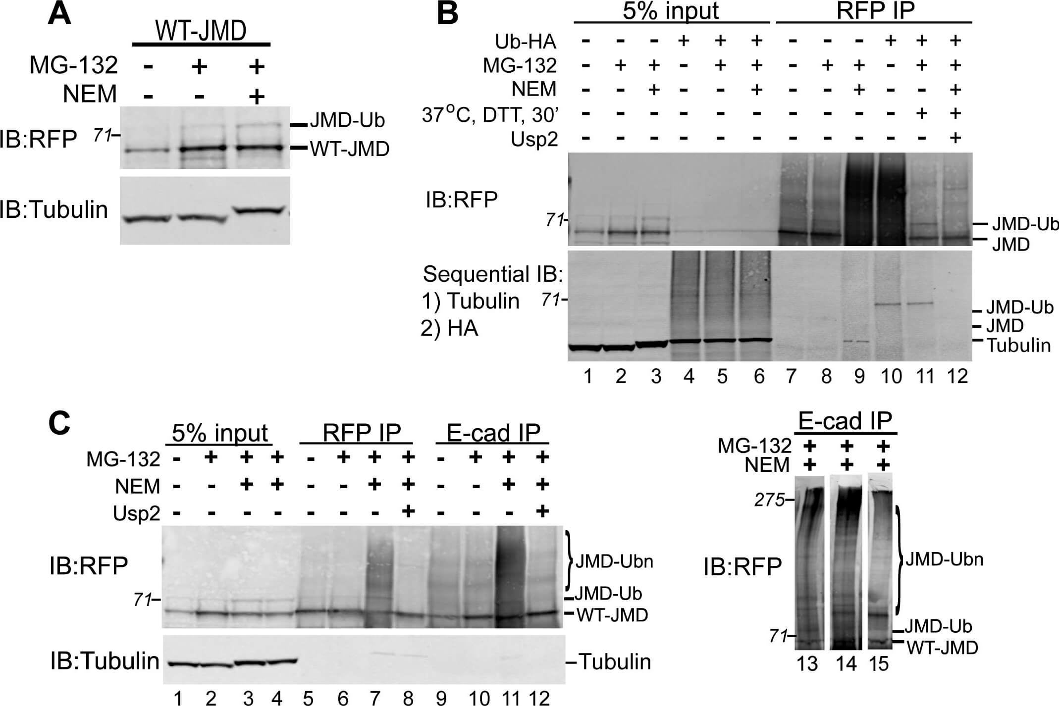
E cadherin JMD is Ubiquitinated.(A) Lysates of WT JMD stable cell lines under normal conditions, upon proteasome inhibition (MG 132), or inhibition of deubiquitinating enzymes (NEM). IB for RFP shows a slower migrating band (JMD Ub) in the presence of MG 132 and NEM. (B) In a separate experiment MDCK cells stably expressing WT JMD were transiently transfected with Ub HA where indicated. Cells were extracted and RFP IP were preformed under normal conditions, upon proteasome inhibition, NEM treatment (to inhibit de ubiquitinating enzymes), addition of the deubiquitinating enzyme Usp2, or mock transfections. DTT was added to the IP after the final was of Protein A beads but prior to the addition of Usp2, where indicated, to neutralize residual NEM. IB were performed for RFP using a rabbit polyclonal antibody, followed by a sequential IB with antibodies specific for:1) tubulin; and 2) HA. The slowest migrating band (marked as JMD Ub) that appears in the presence of NEM is positive for RFP and HA (lanes10,11). HC denotes IgG heavy chain. Number on the side of the gels are the apparent molecular weights of protein standards (*10^3). (C) Extracts from MDCK cells stably expressing WT JMD were immunoprecipitated for RFP and E cadherin under normal conditions, upon proteasome inhibition, NEM treatment (to inhibit de ubiquitinating enzymes), or addition of the de ubiquitinating enzyme Usp2. DTT was added to the IP after the final wash of Protein A beads but prior to the addition of Usp2, where indicated, to neutralize residual NEM. A slow migrating band (lane15) and protein smear (lanes 11,13,14) appear in E cadherin IP in the presence of NEM, and collapse upon incubation with Usp2 (similar to RFP IP). RFP IB in lanes 11,13,14,15 show that the slower migrating band does migrates at different molecular weights indicating variable levels of JMD ubiquitination. Number on the side of the blots are the apparent molecular weights of protein standards (*10^3).
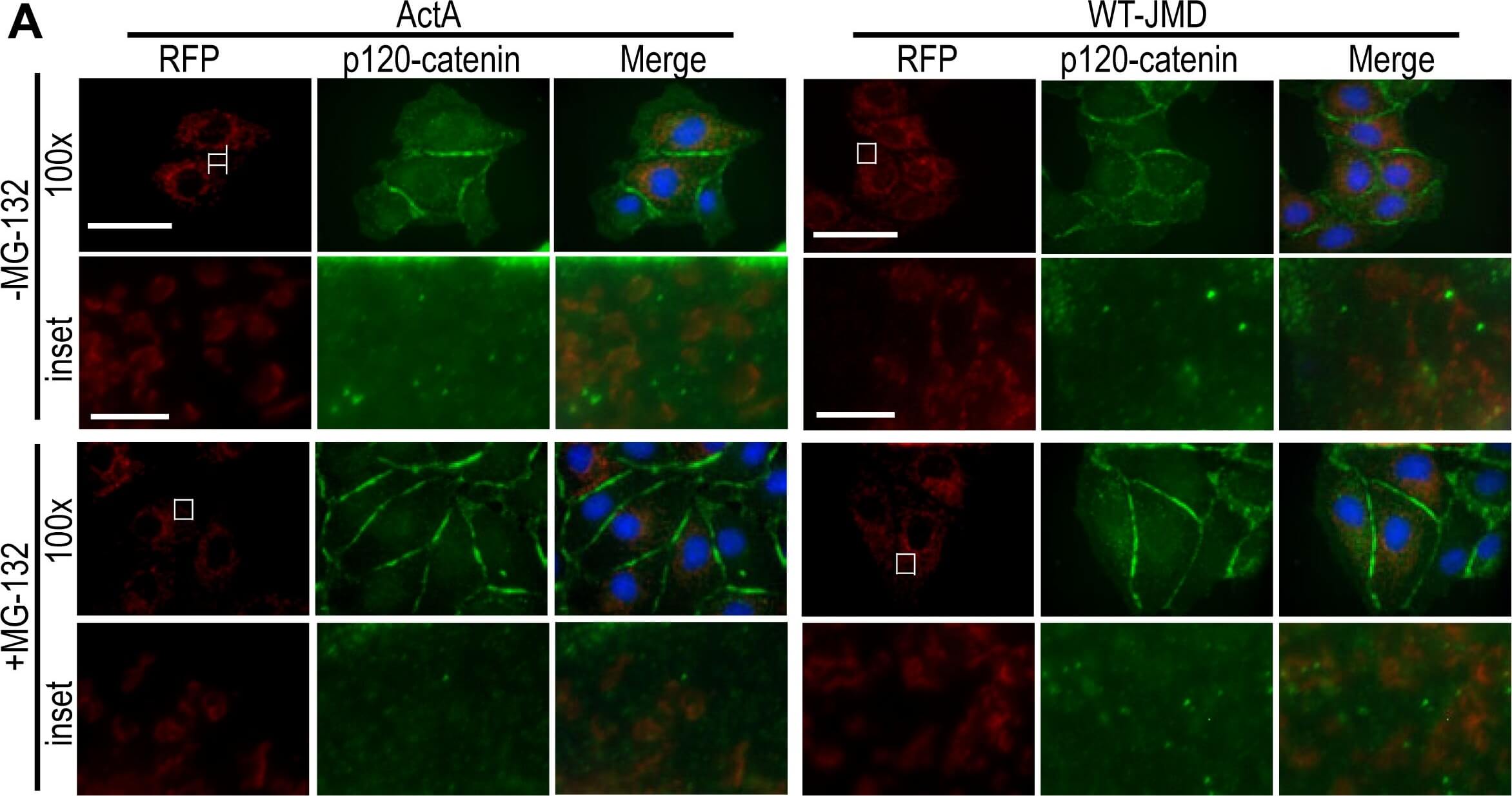
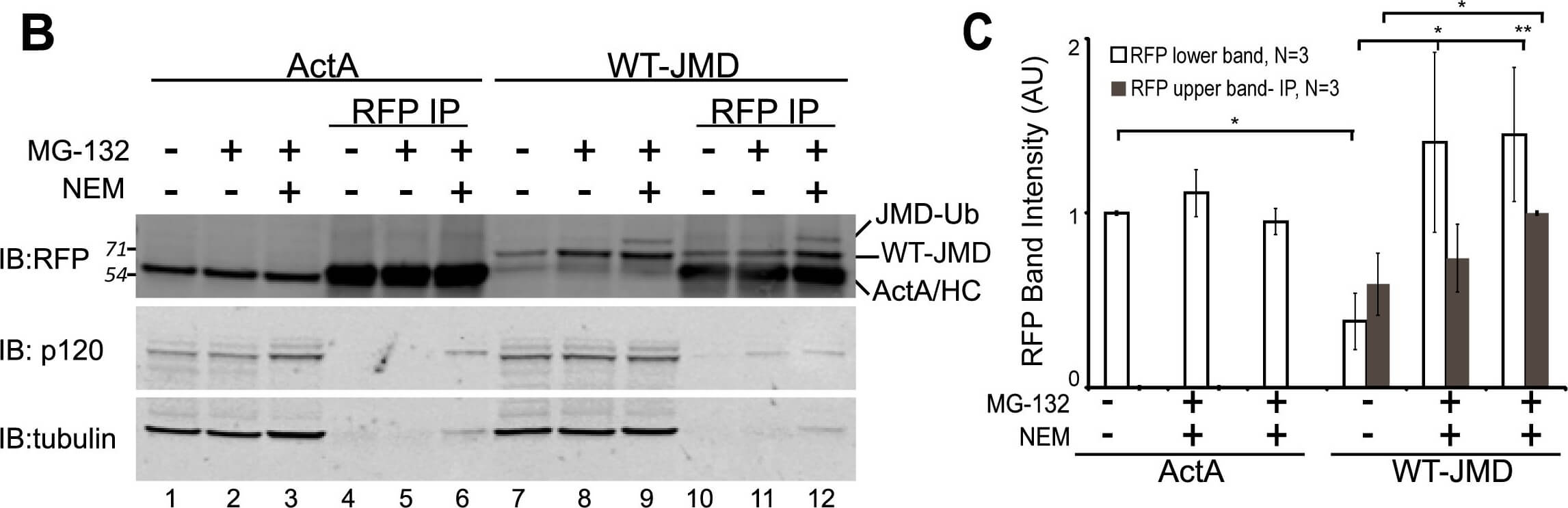
Localization and binding of WT-JMD and p120-catenin.(A) Immunofluorescence of MDCK cells transiently expressing ActA or WT-JMD. Images for RFP (red), p120-catenin (green) and merged are shown separately (100×). Boxed areas are shown as higher magnifications below (RFP-, p120- and merge-inset). All images were from the same experiment and processed identically between cell lines. Scale bar is 25 um in 100× images, and 5 um in insets. (B) Lysates and RFP immunoprecipitates (IP) of ActA and WT-JMD stable cell lines under normal conditions, upon proteasome inhibition, or NEM treatment (to inhibit de-ubiquitinating enzymes). Immunoblots (IB) for RFP show: a slower migrating band (upper band-JMD-Ub) that appears only in WT-JMD cells in the presence of MG-132 and NEM; the band identified as ActA/HC comprises a co-migrating ActA and the IgG heavy chain (HC). Number(s) on the side of the gels are the apparent molecular weights of protein standards (× 10∧3). (C) Quantification of RFP intensities normalized to tubulin in WT-JMD stables cell lines. Data averaged from 3 independent experiments (+/− s.e.m.), and 2 independently cloned stable cell lines; *p?0.05, **p?0.01. Figure provided by CiteAb. Source: PLoS One, PMID: 22693575.
Localization and binding of WT-JMD and p120-catenin.(A) Immunofluorescence of MDCK cells transiently expressing ActA or WT-JMD. Images for RFP (red), p120-catenin (green) and merged are shown separately (100x). Boxed areas are shown as higher magnifications below (RFP-, p120- and merge-inset). All images were from the same experiment and processed identically between cell lines. Scale bar is 25 um in 100x images, and 5 um in insets. (B) Lysates and RFP immunoprecipitates (IP) of ActA and WT-JMD stable cell lines under normal conditions, upon proteasome inhibition, or NEM treatment (to inhibit de-ubiquitinating enzymes). Immunoblots (IB) for RFP show: a slower migrating band (upper band-JMD-Ub) that appears only in WT-JMD cells in the presence of MG-132 and NEM; the band identified as ActA/HC comprises a co-migrating ActA and the IgG heavy chain (HC). Number(s) on the side of the gels are the apparent molecular weights of protein standards (x 10∧3). (C) Quantification of RFP intensities normalized to tubulin in WT-JMD stables cell lines. Data averaged from 3 independent experiments (+/− s.e.m.), and 2 independently cloned stable cell lines; *p<0.05, **p<0.01. Figure provided by CiteAb. Source: PLoS One, PMID: 22693575.
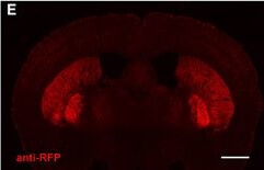
Preserved cell-type specific expression patterns in BAC transgenic mice created with advanced recombineering strategies. (A?D) Anti-2A slice staining in brain slices from DAT-ChETA line 3 mice reveals the distribution of membrane-targeted ChETA protein. Sagittal (A) and coronal (B) sections showing ChR2 expression in midbrain dopamine neurons. (C,D) High magnification images of axon terminal labeling in the dorsal striatum (C) and labeled neurons in the ventral tegmental area (D). (E,F) Anti-RFP slice staining in brain slices from A2A-ChETA line 13 mice showing tdTomato expression in striatopallidal medium spiny neurons (MSNs) in coronal (E) and sagittal (F) sections. Lighter expression can also be detected in putative cortical astrocytes. (G) Anti-2A slice staining in a sagittal brain slice from D1-ChETA line 1 mice showing ChETA expression in striatonigral MSNs. Additional expression is apparent in other brain regions, particularly the dentate gyrus, layer VI cortex, and olfactory bulb. (F) Anti-DARPP32 slice staining to label both striatonigral and striatopallidal MSNs. Scale bars: 1 mm in (A,B,E?H) and 100 μm in (C,D). Figure provided by CiteAb. Source: Front Behav Neurosci, PMID: 24772073.
Preserved cell-type specific expression patterns in BAC transgenic mice created with advanced recombineering strategies. (A?D) Anti-2A slice staining in brain slices from DAT-ChETA line 3 mice reveals the distribution of membrane-targeted ChETA protein. Sagittal (A) and coronal (B) sections showing ChR2 expression in midbrain dopamine neurons. (C,D) High magnification images of axon terminal labeling in the dorsal striatum (C) and labeled neurons in the ventral tegmental area (D). (E,F) Anti-RFP slice staining in brain slices from A2A-ChETA line 13 mice showing tdTomato expression in striatopallidal medium spiny neurons (MSNs) in coronal (E) and sagittal (F) sections. Lighter expression can also be detected in putative cortical astrocytes. (G) Anti-2A slice staining in a sagittal brain slice from D1-ChETA line 1 mice showing ChETA expression in striatonigral MSNs. Additional expression is apparent in other brain regions, particularly the dentate gyrus, layer VI cortex, and olfactory bulb. (F) Anti-DARPP32 slice staining to label both striatonigral and striatopallidal MSNs. Scale bars: 1 mm in (A,B,E?H) and 100 μm in (C,D). Figure provided by CiteAb. Source: Front Behav Neurosci, PMID: 24772073.
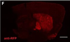
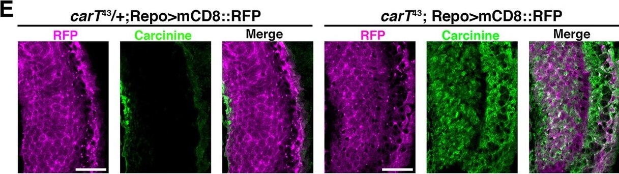
Loss of CarT in photoreceptors increases laminal carcinine.(A) Confocal sections of photoreceptor axonal endings within the lamina stained for the synapse marker Bruchpilot (Brp) and the glia-specific Ebony. (B) Micrographs of cryo-sections from control (OreR) and flies expressing the UAS-Myc-CarT transgene driven by longGMR-Gal4 in the carT43 background. Retina (R), lamina (L), and medulla (M) neuropiles are stained for DNA (magenta) and Myc-tagged CarT (green). Arrow points to Myc staining in photoreceptor axonal endings and arrowhead to distal retina. (C,D) Micrographs of cryo-sections from control (OreR) and mutant flies affecting the histamine?carcinine cycle: carT43, carT43;GMR-Gal4/UAS-Myc-CarT, ebony1, tan1, and HdcMB07212 stained for histamine (C) or carcinine (D). Arrow and arrowhead in D point to carcinine accumulations in the lamina and medulla, respectively. (E) Micrographs from control (carT43/+ ) or carT43?flies expressing a UAS-mCD8::RFP transgene driven by repo-Gal4 and stained for carcinine. (F) Micrographs showing tdTomato fluorescence of the in-frame tdTomato-CarT allele compared with wild type control. DNA staining is shown in green. Arrowheads point to tdTomato signal at R7 and R8 photoreceptor terminals within the medulla. (G) Confocal section of laminal region of a tdTomato-CarT fly stained for glia-specific Ebony. (H) Confocal sections of flies heterozygous for tdTomato-CarT (red) and photoreceptor-specific 3xPax3-eGFP (blue) stained for Brp (green). Sections are parallel to and across photoreceptor axons, respectively. Scale bars are 20 um in A, E, G and H and 50 um in B?D and F.DOI:http://dx.doi.org/10.7554/eLife.10972.007 Figure provided by CiteAb. Source: Elife, PMID: 26653853.
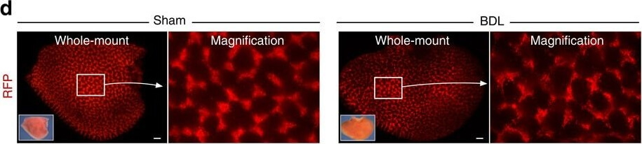
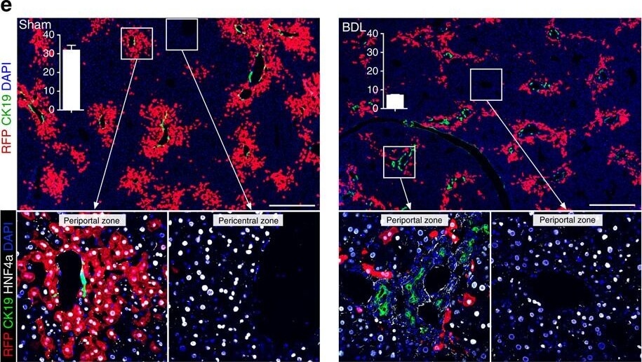
Reduction of pre-labelled PP hepatocytes after injury induced by BDL.(a) Schematic figure showing strategies for PP hepatocytes labelling and injury model induced by BDL. (b) Whole-mount bright view of sham and BDL livers. Scale bars, 2?mm. (c) Sirius red staining of liver sections. Scale bars, 200?μm. (d) Whole-mount fluorescence view of sham and BDL livers. Inserts are bright-field images of the same liver. Scale bars, 500?μm. (e) Immunostaining for RFP, CK19 and HNF4a on sham or BDL liver sections. Boxed regions are magnified in lower panels. Insertions indicate the quantification of the percentage of RFP+ hepatocytes in sham or BDL livers. n=4. Error bars are s.e.m. of the mean. Scale bars, 500?μm. Each image is a representative of four individual samples. Figure provided by CiteAb. Source: Nat Commun, PMID: 27857132.
Reduction of pre-labelled PP hepatocytes after injury induced by BDL.(a) Schematic figure showing strategies for PP hepatocytes labelling and injury model induced by BDL. (b) Whole-mount bright view of sham and BDL livers. Scale bars, 2?mm. (c) Sirius red staining of liver sections. Scale bars, 200?μm. (d) Whole-mount fluorescence view of sham and BDL livers. Inserts are bright-field images of the same liver. Scale bars, 500?μm. (e) Immunostaining for RFP, CK19 and HNF4a on sham or BDL liver sections. Boxed regions are magnified in lower panels. Insertions indicate the quantification of the percentage of RFP+ hepatocytes in sham or BDL livers. n=4. Error bars are s.e.m. of the mean. Scale bars, 500?μm. Each image is a representative of four individual samples. Figure provided by CiteAb. Source: Nat Commun, PMID: 27857132.
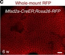
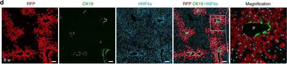
PP hepatocytes labelled by Mfsd2a-CreER.Schematic figures showing (a) our knock-in strategy for Mfsd2a-CreER allele using CRISPR/Cas9 by homologous recombination and (b) genetic lineage tracing strategy for Mfsd2a+ hepatocytes by Cre-LoxP recombination in Mfsd2a+ hepatocytes. (c) Whole-mount fluorescence view of the adult liver from 6-week-old Mfsd2a-CreER;Rosa26-RFP mice. Tamoxifen was induced at 2 days before analysis. Scale bar, 1?mm. (d) Immunostaining for RFP, CK19 and HNF4a on liver sections shows Mfsd2a-expressing hepatocytes in PP zone (P). Scale bars, 100?μm. (e) Immunostaining for RFP, PECAM and 4,6-diamidino-2-phenylindole (DAPI) on liver sections shows Mfsd2a-expressing hepatocytes in the PP zone but not PC zone (*). Scale bars, 100?μm. (f) Isolation of RFP− and RFP+ cells by flow cytometry followed by quantitative RT?PCR (qRT?PCR) analysis for expression of RFP and Mfsd2a. Expression level of genes in the RFP− cells was set as 1. Error bars are s.e.m. of the mean for all the quantification in this study. (g) Expression of PP and PC genes detected by qRT?PCR. The x axis denotes RFP− (black) and RFP+ (red) groups, and the y axis denotes fold induction.. *P<0.05; n=3, two-tailed unpaired t-test. Each immunostaining image is a representative of four individual samples. Figure provided by CiteAb. Source: Nat Commun, PMID: 27857132.
PP hepatocytes labelled by Mfsd2a-CreER.Schematic figures showing (a) our knock-in strategy for Mfsd2a-CreER allele using CRISPR/Cas9 by homologous recombination and (b) genetic lineage tracing strategy for Mfsd2a+ hepatocytes by Cre-LoxP recombination in Mfsd2a+ hepatocytes. (c) Whole-mount fluorescence view of the adult liver from 6-week-old Mfsd2a-CreER;Rosa26-RFP mice. Tamoxifen was induced at 2 days before analysis. Scale bar, 1?mm. (d) Immunostaining for RFP, CK19 and HNF4a on liver sections shows Mfsd2a-expressing hepatocytes in PP zone (P). Scale bars, 100?μm. (e) Immunostaining for RFP, PECAM and 4,6-diamidino-2-phenylindole (DAPI) on liver sections shows Mfsd2a-expressing hepatocytes in the PP zone but not PC zone (*). Scale bars, 100?μm. (f) Isolation of RFP− and RFP+ cells by flow cytometry followed by quantitative RT?PCR (qRT?PCR) analysis for expression of RFP and Mfsd2a. Expression level of genes in the RFP− cells was set as 1. Error bars are s.e.m. of the mean for all the quantification in this study. (g) Expression of PP and PC genes detected by qRT?PCR. The x axis denotes RFP− (black) and RFP+ (red) groups, and the y axis denotes fold induction.. *P<0.05; n=3, two-tailed unpaired t-test. Each immunostaining image is a representative of four individual samples. Figure provided by CiteAb. Source: Nat Commun, PMID: 27857132.
|
|
|
|
E cadherin JMD is Ubiquitinated.(A) Lysates of WT JMD stable cell lines under normal conditions, upon proteasome inhibition (MG 132), or inhibition of deubiquitinating enzymes (NEM). IB for RFP shows a slower migrating band (JMD Ub) in the presence of MG 132 and NEM. (B) In a separate experiment MDCK cells stably expressing WT JMD were transiently transfected with Ub HA where indicated. Cells were extracted and RFP IP were preformed under normal conditions, upon proteasome inhibition, NEM treatment (to inhibit de ubiquitinating enzymes), addition of the deubiquitinating enzyme Usp2, or mock transfections. DTT was added to the IP after the final was of Protein A beads but prior to the addition of Usp2, where indicated, to neutralize residual NEM. IB were performed for RFP using a rabbit polyclonal antibody, followed by a sequential IB with antibodies specific for:1) tubulin; and 2) HA. The slowest migrating band (marked as JMD Ub) that appears in the presence of NEM is positive for RFP and HA (lanes10,11). HC denotes IgG heavy chain. Number on the side of the gels are the apparent molecular weights of protein standards (*10^3). (C) Extracts from MDCK cells stably expressing WT JMD were immunoprecipitated for RFP and E cadherin under normal conditions, upon proteasome inhibition, NEM treatment (to inhibit de ubiquitinating enzymes), or addition of the de ubiquitinating enzyme Usp2. DTT was added to the IP after the final wash of Protein A beads but prior to the addition of Usp2, where indicated, to neutralize residual NEM. A slow migrating band (lane15) and protein smear (lanes 11,13,14) appear in E cadherin IP in the presence of NEM, and collapse upon incubation with Usp2 (similar to RFP IP). RFP IB in lanes 11,13,14,15 show that the slower migrating band does migrates at different molecular weights indicating variable levels of JMD ubiquitination. Number on the side of the blots are the apparent molecular weights of protein standards (*10^3).
|
|
|
| メーカー |
品番 |
包装 |
|
RKL
|
600-401-379
|
100 UG
|
※表示価格について
|

