|
※サムネイル画像をクリックすると拡大画像が表示されます。
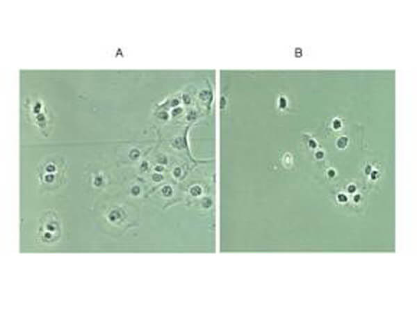
Rockland Immunochemical's Rabbit anti-p65 NLS was used at a 1:200 dilution to detect p65 in (A) control DU145 cells and (B) TNF stimulated DU145 cells.? Although DU145 show relatively high basal levels of nuclear p65 staining, significant enhancement of nuclear staining is seen in panel B as evidence of translocation and availability of the NLS to be bound by the antibody.? Cultured cells shown above were formalin-fixed.? Tissue staining (not shown) were formalin-fixed, paraffin embedded followed by citrate retrieval.? Blocking and hybridization included 5% NGS.
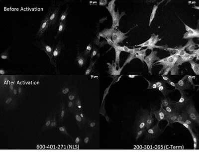
Anti-p65 NLS Antibody and Anti NFkB monoclonal antibody ? Immunocytochemistry. Tissue: Human Fibroblasts. Top two: Before activation. Bottom two: After activation with poly IC. Left: 600-401-271: anti-p65 NLS specific lot 18372. Right: 200-301-065: Monoclonal anti-NFkB p65 antibody C-Term. The two antibodies that are shown target different regions of the p65 protein. The different staining patterns are thought to correspond with different functional regions of the protein.
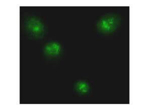
Immunofluorescence Microscopy of Anti-p65 NLS Antibody. Cells: TNF stimulated DU145 cells - fixed. Antibody: Rabbit anti-p65 NLS was used at a 1:200 dilution to detect p65. Image shown is at a 1:400 magnification.
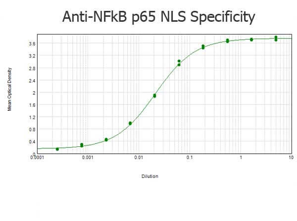
ELISA results of purified Rabbit anti-NFkB p65 NLS Specific Antibody tested against BSA-conjugated peptide of immunizing peptide. Each well was coated in duplicate with 0.1μg of conjugate. The starting dilution of antibody was 5μg/ml and the X-axis represents the Log10 of a 3-fold dilution. This titration is a 4-parameter curve fit where the IC50 is defined as the titer of the antibody. Assay performed using 3% fish gel, Goat anti-Rabbit IgG Antibody Peroxidase Conjugated (Min X Bv Ch Gt GP Ham Hs Hu Ms Rt & Sh Serum Proteins) (p/n 611-103-122) and TMB ELISA Peroxidase Substrate (p/n TMBE-1000).
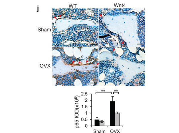
(j) Immunostaining and quantification of active p65 in trabecular bone cells and surrounding bone marrow cells in WT and Wnt4 mice after OVX or sham operation. Scale bars, 30 μm. IOD, integral optical density.?n?= 8 for sham groups;?n?= 12 for OVX groups. *P?< 0.05, **?P?< 0.01, unpaired two-tailed t-test. Fig 2. PMID:?25108526
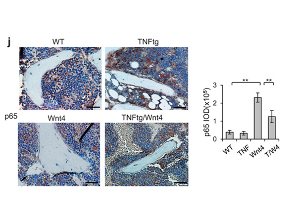
(j) Immunostaining with anti-active p65 and quantification of NF-κB activity surrounding the trabecular bone in WT, Wnt4, TNFtg and TNFtg/Wnt4 mice. Scale bars, 40μm. TNF, TNFtg mice; T/W4, TNFtg/Wnt4 mice.?n?= 6 per group for WT and WNT4 mice;?n?= 8 per group for TNFtg and TNFtg/Wnt mice. *P?< 0.05, **?P?< 0.01, unpaired two-tailed t-test. Fig 3. PMID:?25108526
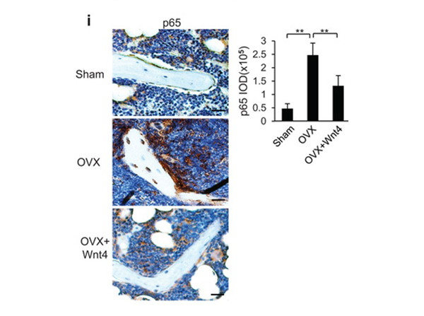
?(i) Immunostaining with anti-active p65 and quantification of NF-κB activity surrounding the trabecular bones from mice after sham operation, OVX and OVX with rWnt4 injection. Scale bars, 30 μm. Fig 6. PMID:?25108526
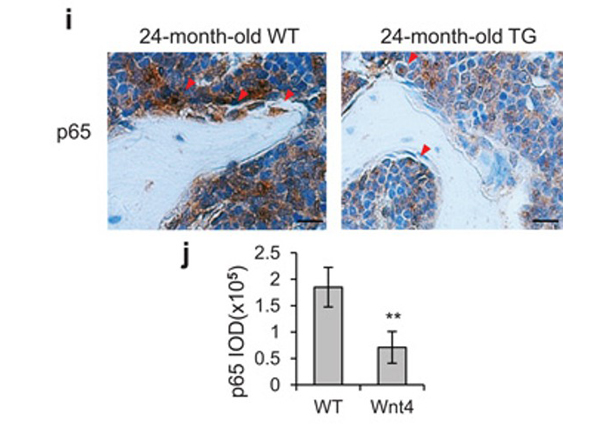
(i) Immunostaining with anti-active p65 and quantification of NF-κB activity surrounding the trabecular bones from 24-months-old WT and Wnt4 mice. Scale bars, 25 μm. n?= 12 mice per group. *P?< 0.05, **?P?< 0.01, unpaired two-tailed t-test. Fig 4. PMID:?25108526
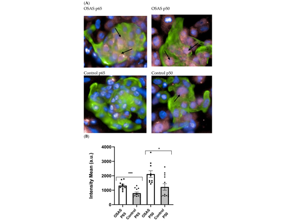
NF-κB is activated by stimulation with sera from OSAS patients. (A) A representative picture of a cluster of cells is presented. Cardiomyocytes are identified by staining with anti-cardiac troponin (green). NF-κB subunits: Anti p50 or anti p65 (pink). Arrows point to nuclei expressing p50 or p65. (B) The total average intensity of NF-κB subunits p50 and p65 staining was measured in the nuclei of cardiomyocytes. The cells were incubated each time with 12 different OSAS sera and compared to 10 different control sera (5%). The results presented are an average of 3 separate experiments done in duplicates, showing a significant increase of nuclear p50 and p65 following incubation with OSAS sera. Squares and circles represent individual sera. Statistical significance was determined by the Student’s?t-test comparing cells incubated with control or OSAS sera (p50?p?= 0.01, p65?p?= 0.001). *?p?< 0.05, ***?p?< 0.01. Fig 2. PMID:?34768848
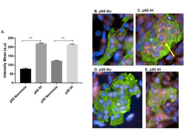
Increase in nuclear NF-κB sub-units p65 and p50 expression in hESC-CMs following IH: (A) quantification of the NF-κB subunits p65 and p50 expression in the nucleus following normoxia or IH only on CMs. Normoxia (21% O2, 37 °C), intermittent hypoxia (IH) (1% O2, 37 °C) for 12 h (60 Cycles). Results are averages of 3 separate experiments performed in 10 replicates Student’s t-test ***?p?< 0.001, n = 39,852; (B) immunostaining of p65, p50 and cardiac troponin T (cardiomyocytes specific marker) following normoxia or IH. Differentiated CMs were detected with antibodies to cardiac troponin T (green) and p65 or p50 (pink), followed by the appropriate secondary antibody. Nuclei were stained with DAPI (blue); (B) normoxia conditions and p65; (C) IH conditions and p65; (D) normoxia conditions and p50; and (E) IH conditions and p50. 40× magnification. Fig 3. PMID:?36142186
|
|
|
|
Rockland Immunochemical's Rabbit anti-p65 NLS was used at a 1:200 dilution to detect p65 in (A) control DU145 cells and (B) TNF stimulated DU145 cells.? Although DU145 show relatively high basal levels of nuclear p65 staining, significant enhancement of nuclear staining is seen in panel B as evidence of translocation and availability of the NLS to be bound by the antibody.? Cultured cells shown above were formalin-fixed.? Tissue staining (not shown) were formalin-fixed, paraffin embedded followed by citrate retrieval.? Blocking and hybridization included 5% NGS.
|
|
| 別品名 |
rabbit anti-NFKB p65 antibody, rabbit anti-p65 antibody, rabbit anti-NLS specific antibody, nuclear localization sequence, NFKB, NFkβ, NF-kB, NF-kappaB, NFkappaB, Transcription factor p65, Nuclear factor NF-kappa-B p65 subunit, Nuclear factor of kappa light polypeptide gene enhancer in B-cells 3, RELA, NFKB3
|
| 交差種 |
Human
|
| 適用 |
Western Blot
Enzyme Linked Immunosorbent Assay
Immuno Fluorescence
|
| 免疫動物 |
Rabbit
|
| 標識物 |
Unlabeled
|
| 精製度 |
Affinity Purified
|
| GENE ID |
5970
|
| Accession No.(Gene/Protein) |
223468676, Q04206
|
| Gene Symbol |
RELA
|
| 参考文献 |
[[Cosmo Reference Collection]][Pub Med ID]16644679,25108526,29637625,30244868,34768848,36142186
|
| [注意事項] |
濃度はロットによって異なる可能性があります。メーカーDS及びCoAからご確認ください。
|
|
| メーカー |
品番 |
包装 |
|
RKL
|
600-401-271
|
100 UG
|
※表示価格について
| 当社在庫 |
なし
|
| 納期目安 |
約10日
|
| 保存温度 |
-20℃
|
|
※当社では商品情報の適切な管理に努めておりますが、表示される法規制情報は最新でない可能性があります。
また法規制情報の表示が無いものは、必ずしも法規制に非該当であることを示すものではありません。
商品のお届け前に最新の製品法規制情報をお求めの際はこちらへお問い合わせください。
|
※当社取り扱いの試薬・機器製品および受託サービス・創薬支援サービス(納品物、解析データ等)は、研究用としてのみ販売しております。
人や動物の医療用・臨床診断用・食品用としては、使用しないように、十分ご注意ください。
法規制欄に体外診断用医薬品と記載のものは除きます。
|
|
※リンク先での文献等のダウンロードに際しましては、掲載元の規約遵守をお願いします。
|
|
※CAS Registry Numbers have not been verified by CAS and may be inaccurate.
|


