|
※サムネイル画像をクリックすると拡大画像が表示されます。
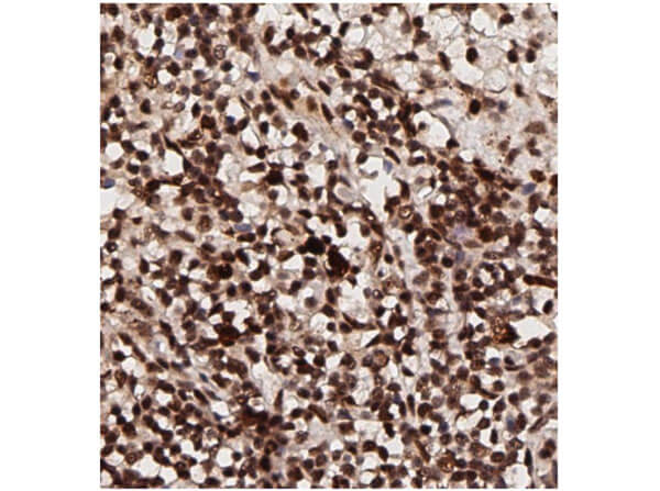
Immunohistochemistry of Rabbit Anti-AKT pT308 Antibody. Tissue: human breast tissue (lymph nodes). Antigen retrieval: HIER using citrate buffer for 20 minutes. Fixative: None. Primary Antibody: Anti-AKT phosphoT308 at 1:200 for 30 minutes at RT. Secondary Antibody: Anti-Rabbit Poly-HRP-IgG Ready to Use for 8 minutes at RT. Counterstain: Hematoxylin. Substrate: DAB. Analysis: Strong staining in nucleus. May be suitable with more dilutions.
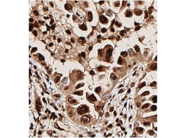
Immunohistochemistry of Rabbit Anti-AKT pT308 Antibody. Tissue: human lung tissue. Antigen retrieval: HIER using citrate buffer for 20 minutes. Fixative: None. Primary Antibody: Anti-AKT phosphoT308 at 1:200 for 30 minutes at RT. Secondary Antibody: Anti-Rabbit Poly-HRP-IgG Ready to Use for 8 minutes at RT. Counterstain: Hematoxylin. Substrate: DAB. Analysis: Strong staining in nucleus. May be suitable with more dilutions.
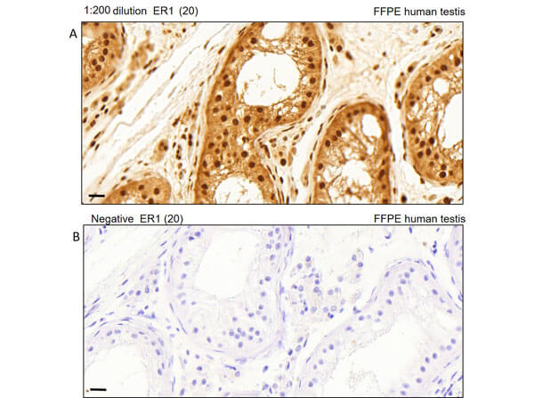
Immunohistochemistry of Rabbit Anti-AKT pT308 Antibody. Tissue: human testis tissue. Antigen retrieval: Heat induced antigen retrieval was performed using Leica Bond Epitope Retrieval Buffer 1 (Citrate solution, pH6.0) for 20 minutes. Fixative: None. Primary Antibody: (A). Anti-AKTpT308 at 1:200 for 30 minutes at RT. (B). Negative control. Secondary Antibody: Anti-Rabbit Poly-HRP-IgG Ready to Use for 8 minutes at RT. Counterstain: Hematoxylin. Substrate: DAB. Analysis: Cells in seminiferous ducts and Leydig cells show moderate cytoplasmic staining.
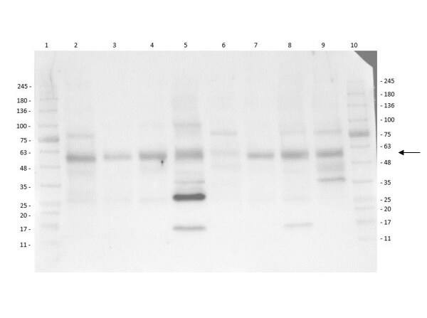
Multi-Lysate Western Blot of Rabbit Anti-AKT pT308 Antibody. Lane 1: Opal Pre-stained Molecular Weight Marker (p/n MB-210-0500). Lane 2: Human Spleen Lysate. Lane 3: Hu Small Intestine Lysate. Lane 4: Hu Placenta Lysate. Lane 5: Hu Skeletal Muscle Lysate. Lane 6: Hu Brain Cerebellum Lysate. Lane 7: Hu Lung Lysate. Lane 8: Hu Tonsil Lysate. Lane 9: Hu Thymus Lysate. Lane 10: Opal Pre-stained Molecular Weight Marker (p/n Mb-210-0500). Primary Antibody: Anti-AKT pT308 at 1:1000 overnight at 2-8°C. Secondary Antibody: Goat Anti-Rabbit IgG HRP (p/n 611-103-122) at 1:40,000 at RT for 60mins. Block: BlockOut Buffer (p/n MB-073). Predicted MW: ~55kDa. Observed MW: ~28, ~58kDa. Notes: Ubiquitous.
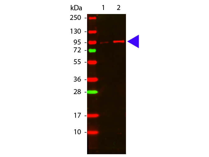
Western Blot of Rabbit anti-Akt phospho T308 antibody. Lane 1: GST tagged AKT1 un-active recombinant protein. Lane 2: GST tagged AKT1 active recombinant protein. Load: 50 ng per lane. Primary antibody: Akt phospho T308 antibody at 1:1,000 for overnight at 4°C. Secondary antibody: DyLight? 649 rabbit secondary antibody at 1:20,000 for 30 min at RT. Block: MB-070 for 30 min at RT. Other band(s): none.
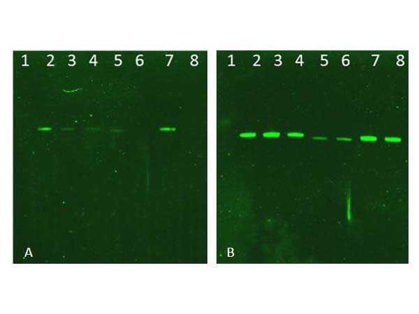
Western Blot of Rabbit AKT Antibodies. Lane 1: NIR MW protein ladder. Lane 2: AKT1, recombinant: 009-001-P21. Lane 3: AKT1, phosphatase-treated: 009-001-I51. Lane 4: AKT1, mutant T308A/S473A: 009-001-P22. Lane 5: AKT2, recombinant: 009-001-P23. Lane 6: AKT2, phosphatase-treated: 009-001-E71. Lane 7: AKT3, recombinant: 009-001-P24. Lane 8: AKT3, phosphatase-treated: 009-001-E75. Load: 50ng per lane. Blot A: 600-401-269 Anti-Akt pT308 used at 1:2270, Blot B: 100-401-401 Anti-Akt used 1:1000.
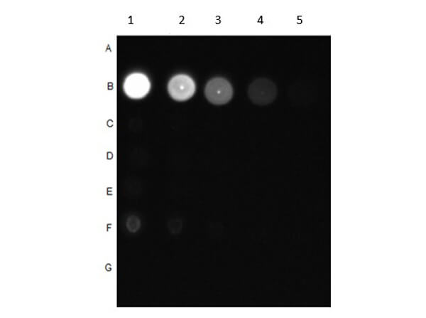
Dot Blot of Rabbit Anti-AKT pT308 Antibody. Dilutions in Columns: (1) 100ng, (2) 33.33ng, (3) 11.11ng, (4) 3.7ng, (5) 1.23ng. Tested BSA Peptide Reactivity in Rows: (A) AKT1-BSA, (B) AKT1 pT308-BSA, (C) AKT1 S473-BSA, (D) AKT1 pS473-BSA, (E) CDC27 T244-BSA, (F) CDC27 pT244-BSA, (G) BSA control. Primary Antibody: Anti-AKT pT308 at 1μg/mL overnight at 2-8°C. Secondary Antibody: Goat anti-Rabbit IgG HRP (p/n 611-103-122) at 1:70,000 at RT for 30mins. Block: BlockOut Buffer (p/n MB-073).
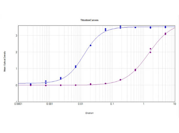
ELISA Results of Rabbit Anti-AKT pT308 Antibody tested against BSA-conjugated non-phospho [purple] and phospho [blue] forms of immunizing peptide. Each well was coated in duplicate with either 0.1μg of conjugate. The working dilution is 1:81,300. The starting dilution of antibody was 5μg/ml and the X-axis represents the Log10 of a 3-fold dilution. This titration is a 4-parameter curve fit where the IC50 is defined as the titer of the antibody. Assay performed using HRP conjugate stabilizer (p/n MB-076), Goat Anti-Rabbit HRP conjugated (p/n 611-103-122) and TMB substrate (p/n TMBE-1000).
|
|
|
|
Immunohistochemistry of Rabbit Anti-AKT pT308 Antibody. Tissue: human breast tissue (lymph nodes). Antigen retrieval: HIER using citrate buffer for 20 minutes. Fixative: None. Primary Antibody: Anti-AKT phosphoT308 at 1:200 for 30 minutes at RT. Secondary Antibody: Anti-Rabbit Poly-HRP-IgG Ready to Use for 8 minutes at RT. Counterstain: Hematoxylin. Substrate: DAB. Analysis: Strong staining in nucleus. May be suitable with more dilutions.
|
|
| 別品名 |
rabbit anti-AKT pT308 Antibody, AKT1 phospho T308, RAC-PK-alpha, Protein kinase B, PKB, C-AKT, RAC-alpha serine/threonine-protein kinase, Proto-oncogene c-Akt, AKT1, AKT 1, AKT-1
|
| 交差種 |
Human
Mouse
Rat
|
| 適用 |
Western Blot
Enzyme Linked Immunosorbent Assay
Immunohistochemistry
Dot Blot
|
| 免疫動物 |
Rabbit
|
| 抗原部位 |
a.a.302-311
|
| 標識物 |
Unlabeled
|
| 精製度 |
Affinity Purified
|
| 翻訳後修飾 |
リン酸化
|
| GENE ID |
207
|
| Accession No.(Gene/Protein) |
62241011, P31749
|
| Gene Symbol |
AKT1
|
| 参考文献 |
Lawlor, M. A. and Alessi, D.R. (2001). PKB/AKT: a key mediator of cell proliferation, survival and insulin responses. J. Cell Science 114:2903-2910. Alessi, D. R. (2001). Discovery of PDK1, one of the missing links in insulin signal transduction. Biochem. Soc. Trans. 29,1 -14. Hedjazifar,S., Jenndahl,L.E., Shimokawa,H. and Baeckstrom,D. (2005) PKB mediates c-erbB2-induced epithelial beta1 integrin conformational inactivation through Rho-independent F-actin rearrangements. Exp. Cell Res. 307 (1), 259-275. Saji,M., Vasko,V., Kada,F., Allbritton,E.H., Burman,K.D. and Ringel,M.D. (2005) Akt1 contains a functional leucine-rich nuclear export sequence. Biochem. Biophys. Res. Commun. 332 (1), 167-173. Sutherland,B.W., Kucab,J., Wu,J., Lee,C., Cheang,M.C., Yorida,E., Turbin,D., Dedhar,S., Nelson,C., Pollak,M., Leighton Grimes,H., Miller,K., Badve,S., Huntsman,D., Blake-Gilks,C., Chen,M., Pallen,C.J. and Dunn,S.E. (2005) Akt phosphorylates the Y-box binding protein 1 at Ser102 located in the cold shock domain and affects the anchorage-independent growth of breast cancer cells. Oncogene 24 (26), 4281-4292.
|
| [注意事項] |
濃度はロットによって異なる可能性があります。メーカーDS及びCoAからご確認ください。
|
|
| メーカー |
品番 |
包装 |
|
RKL
|
600-401-269
|
100 UG
|
※表示価格について
| 当社在庫 |
なし
|
| 納期目安 |
約10日
|
| 保存温度 |
-20℃
|
|
