|
※サムネイル画像をクリックすると拡大画像が表示されます。
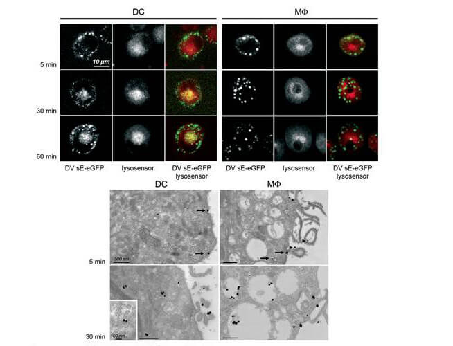
Immuno-microscopy of Rabbit anti-GFP antibody. Monocyte derived dendritic cells and dermal macrophages were challenged and directly visualized with eGFP labeled Dengue virus to localize sequestration of virus particles in the different cells (upper). The location of the GFP was confirmed by TEM (lower magnified view) using Rockland rabbit anti GFP Primary antibody (1:200) and a gold labeled secondary antibody. As referenced in: Kwan W-H, Navarro-Sanchez E, Dumortier H, Decossas M, Vachon H, et al. (2008) Dermal-Type Macrophages Expressing CD209/DC-SIGN Show Inherent Resistance to Dengue Virus Growth. PLoS Negl Trop Dis 2(10): e311. doi:10.1371/journal.pntd.0000311
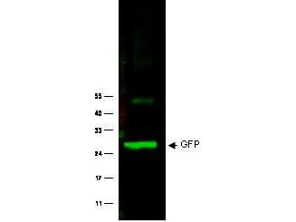
Western Blot of Rabbit anti-GFP antibody. Lane 1: Wild type GFP (0.1 μg) was used to spike HeLa whole cell lysate. Lane 2: none. Load: 30 μg per lane. Primary antibody: GFP antibody at 1:1000 for overnight at 4°C. Secondary antibody: IRDye800? Goat-a-Rabbit IgG [H&L] MX10 (611-132-122) at 1:10,000 for 45 min at RT. Block: 5% BLOTTO in PBS overnight at 4°C. Predicted/Observed size: 27 kDa for epitope tag GFP. Other band(s): none.
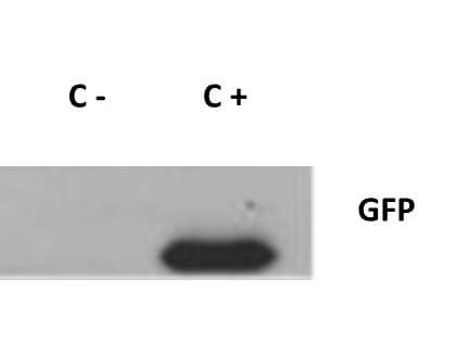
Western Blot of Rabbit anti-GFP antibody. Lane 1: 293FT cells transfected with CDK4 dominant negative (C-). Lane 2: 293FT cells poitive control (C+). Load: 25 μg per lane. Primary antibody: GFP antibody at 1:400 for overnight at 4°C. Secondary antibody: IRDye800? rabbit secondary antibody at 1:10,000 for 45 min at RT. Block: 5% BLOTTO overnight at 4°C. Predicted/Observed size: 27 kDa for GFP.
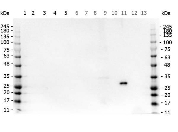
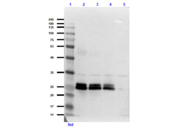
Western Blot of Rabbit anti-GFP antibody. Marker: Opal Pre-stained ladder (p/n MB-210-0500). Lane 1: HEK293 lysate (p/n W09-000-365). Lane 2: HeLa Lysate (p/n W09-000-364). Lane 3: CHO/K1 Lysate (p/n W07-000-357). Lane 4: MDA-MB-231 (p/n W09-001-GK6). Lane 5: A431 Lysate (p/n W09-000-361). Lane 6: Jurkat Lysate (p/n W09-001-370). Lane 7: NIH/3T3 Lysate (p/n W10-000-358). Lane 8: E-coli HCP Control (p/n 000-001-J08). Lane 9: FLAG Positive Control Lysate (p/n W00-001-383). Lane 10: Red Fluorescent Protein (p/n 000-001-379). Lane 11: Green Fluorescent Protein (p/n 000-001-215). Lane 12: Glutathione-S-Transferase Protein (p/n 000-001-200). Lane 13: Maltose Binding Protein (p/n 000-001-385). Load: 10 μg of lysate or 50ng of purified protein per lane. Primary antibody: GFP antibody at 1ug/mL overnight at 4C. Secondary antibody: Peroxidase rabbit secondary antibody (p/n 611-103-122) at 1:30,000 for 60 min at RT. Blocking Buffer: 1% Casein-TTBS (p/n MB-082) for 30 min at RT. Predicted/Observed size: 30 kDa for GFP.
Western Blot of Rabbit Anti-GFP Antibody. Lane 1: Opal Prestained Molecular Weight Ladder (p/n MB-210-0500). Lane 2: GFP (p/n 000-001-215) / HeLa Lysate (p/n W09-000-364) [0.1μg/10.0μg]. Lane 3: GFP (p/n 000-001-215) / HeLa Lysate (p/n W09-000-364) [0.05μg/10.0μg]. Lane 4: GFP (p/n 000-001-215) / HeLa Lysate (p/n W09-000-364) [0.03μg/10.0μg]. Lane 5: HeLa Lysate (p/n W09-000-364) [10.0μg]. Primary Antibody: Rabbit Anti-GFP Antibody at 1.0μg/mL overnight at 2-8°C. Secondary Antibody: Goat Anti-Rabbit IgG MX 10 Peroxidase (p/n 611-103-122) at 1:70,000 for 30mins at RT. Block: Blocking Buffer for Fluorescent Western Blotting (p/n MB-070) for 60 mins at RT. Expect: ~27kDa.
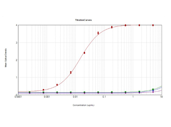
ELISA results of purified Rabbit Anti-GFP Antibody. Each well was coated in 10 μg of antigen GFP [Red Line], human IgG [Green Line], Mouse IgG [Blue Line], and Rat IgG [Purple Line]. The starting dilution of antibody was 5 μg/mL and the X-axis represents the Log10 of a 3-fold dilution. The titer is 1:67,700. This titration is a 4-parameter curve fit where the IC50 is defined as the titer of the antibody. Assay performed using 1% Fish Gel, TMB Substrate (p/n TMB-1000), and Goat Anti-Rabbit IgG Antibody HRP (p/n 611-103-122).
|
|
|
|
Immuno-microscopy of Rabbit anti-GFP antibody. Monocyte derived dendritic cells and dermal macrophages were challenged and directly visualized with eGFP labeled Dengue virus to localize sequestration of virus particles in the different cells (upper). The location of the GFP was confirmed by TEM (lower magnified view) using Rockland rabbit anti GFP Primary antibody (1:200) and a gold labeled secondary antibody. As referenced in: Kwan W-H, Navarro-Sanchez E, Dumortier H, Decossas M, Vachon H, et al. (2008) Dermal-Type Macrophages Expressing CD209/DC-SIGN Show Inherent Resistance to Dengue Virus Growth. PLoS Negl Trop Dis 2(10): e311. doi:10.1371/journal.pntd.0000311
|
|
| 別品名 |
rabbit anti-GFP antibody, Green Fluorescent Protein, GFP antibody, Green Fluorescent Protein antibody, EGFP, enhanced Green Fluorescent Protein, Aequorea victoria, Jellyfish
|
| 適用 |
Western Blot
Enzyme Linked Immunosorbent Assay
|
| 免疫動物 |
Rabbit
|
| 標識物 |
Unlabeled
|
| 精製度 |
Affinity Purified
|
| Accession No.(Gene/Protein) |
P42212
|
| Tag情報 |
GFP
|
| 参考文献 |
[Pub Med ID]31067451
|
| [注意事項] |
濃度はロットによって異なる可能性があります。メーカーDS及びCoAからご確認ください。
|
|
| メーカー |
品番 |
包装 |
|
RKL
|
600-401-215
|
100 UG
|
※表示価格について
| 当社在庫 |
なし
|
| 納期目安 |
約10日
|
| 保存温度 |
-20℃
|
|
※当社では商品情報の適切な管理に努めておりますが、表示される法規制情報は最新でない可能性があります。
また法規制情報の表示が無いものは、必ずしも法規制に非該当であることを示すものではありません。
商品のお届け前に最新の製品法規制情報をお求めの際はこちらへお問い合わせください。
|
※当社取り扱いの試薬・機器製品および受託サービス・創薬支援サービス(納品物、解析データ等)は、研究用としてのみ販売しております。
人や動物の医療用・臨床診断用・食品用としては、使用しないように、十分ご注意ください。
法規制欄に体外診断用医薬品と記載のものは除きます。
|
|
※リンク先での文献等のダウンロードに際しましては、掲載元の規約遵守をお願いします。
|
|
※CAS Registry Numbers have not been verified by CAS and may be inaccurate.
|
