|
※サムネイル画像をクリックすると拡大画像が表示されます。
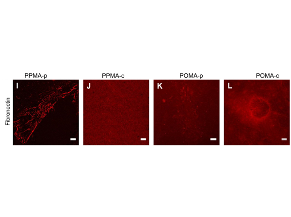
Immunofluorescence of Anti-Fibronectin Antibody. Representative images of isolated EC cultured on various MA-coated surfaces after 24 h exposure to 0.5 dyn/cm2 immunofluorescence-labelled for Fibronectin. A strong rearrangement of the initial Fibronectin layer into coarse fibrils (under venous shear stress) (Fig. 3I) with severe displacements of Fibronectin occurred on PPMA-p. Only slight Fibronectin reorganization into fine fibrils (PPMA-c Fig. 3J) or no Fibronectin reorganization at all (POMA-p and POMA-c Fig. 3K and L) were observed as expected from the higher Fibronectin anchorage strength to these latter substrates in comparison to PPMA-p. These findings are in line with earlier results at static cell culture conditions of isolated EC [4e6] showing the dependence of adhesion and stress fibre patterns on the matrix anchorage to the polymer surface, which were now attenuated by the application of shear stress.Scale bar: 10 mm. Fig. 3. PMID: 22154622.
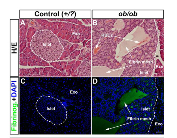
Immunohistochemistry of Anti-Fibronectin Antibody. Immunohistochemical assessment of proteins involved in blood coagulation in ob/ob pancreas. (A,B) Hematoxylin/Eosin staining of an islet from a lean control (A) and a ob/ob (B) pancreas at 52 weeks. Note the accumulation of RBCs (white arrows in (B). (C,D) Consecutive sections to (A,B) stained for Fibrinogen (green) and DAPI (Blue) indicating the presence of a fibrin mesh within the areas of the lesions (white arrows in (D) compare with (B)). Scale bar in (D) is 50μm. Figure 6. PMID: 27713548.
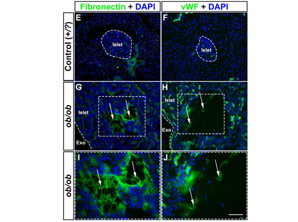
Immunohistochemistry of Anti-Fibronectin Antibody. Immunohistochemical assessment of proteins involved in blood coagulation in ob/ob pancreas. (E?J) Photomicrographs of representative pancreatic cryosections from lean control (E,F) and ob/ob (G,H) pancreas at 52 weeks of age labeled for Fibronectin (Green E,G) and von Willebrand Factor (Green, F,H) together with DAPI (blue). Areas enclosed by a broken line in (G,H) corresponds to (I,J) respectively. The areas in the lesions positive for Fibronectin and von Willebrand factor are not associated with any nucleated cells. Abbreviations; vWF, von Willebrand Factor; Exo, Exocrine tissue. Scale bar in (J) is 92μm in (E?H) and 50μm in (I,J). Figure 6. PMID: 27713548.
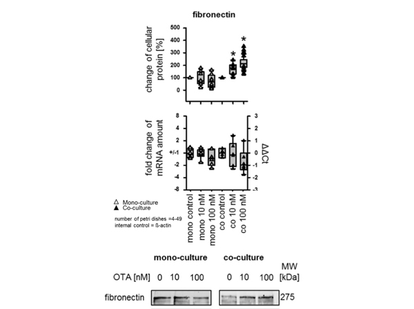
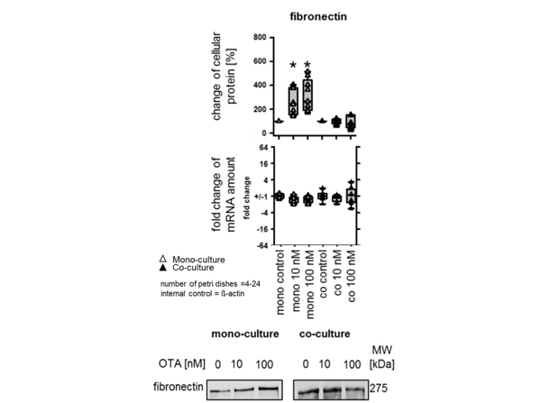
Western Blot of Anti-Fibronectin Antibody. Impact of OTA on the cellular protein and mRNA amount in renal epithelial cells [NRK-52E]. OTA effect on cellular fibronectin protein amount and mRNA abundance in NRK-52E under mono- and co-culture conditions. Representative Western blots of proteins isolated from cells exposed to OTA. * indicates significant difference compared with the control group.In the presence of fibroblasts, exposure to 10 nM OTA led to an increase of fibronectin protein amount. Incubation with 100 nM OTA led to an increase of fibronectin protein amount. (Fig. 3 only fibronectin displayed). Fig. 3. PMID: 31415839.
Western Blot of Anti-Fibronectin Antibody.Impact of OTA on cellular protein and mRNA amount in fibroblasts [NRK-49F]. OTA effect on fibronectin protein amount and mRNA abundance in NRK-49F under mono and co-culture conditions. Representative Western blots of proteins isolated from cells exposed to OTA. * indicates significant difference compared with the control group. Exposure of fibroblasts in monoculture to 10 or 100 nM OTA caused an increase of fibronectin protein amount. Fig. 4. PMID: 31415839.
|
|
|
|
Immunofluorescence of Anti-Fibronectin Antibody. Representative images of isolated EC cultured on various MA-coated surfaces after 24 h exposure to 0.5 dyn/cm2 immunofluorescence-labelled for Fibronectin. A strong rearrangement of the initial Fibronectin layer into coarse fibrils (under venous shear stress) (Fig. 3I) with severe displacements of Fibronectin occurred on PPMA-p. Only slight Fibronectin reorganization into fine fibrils (PPMA-c Fig. 3J) or no Fibronectin reorganization at all (POMA-p and POMA-c Fig. 3K and L) were observed as expected from the higher Fibronectin anchorage strength to these latter substrates in comparison to PPMA-p. These findings are in line with earlier results at static cell culture conditions of isolated EC [4e6] showing the dependence of adhesion and stress fibre patterns on the matrix anchorage to the polymer surface, which were now attenuated by the application of shear stress.Scale bar: 10 mm. Fig. 3. PMID: 22154622.
|
|
| 別品名 |
rabbit anti-Fibronectin antibody, FN1, FN, Cold-insoluble globulin, CIG, Anastellin, Ugl-Y1, Ugl-Y2, Ugl-Y3
|
| 交差種 |
Human
|
| 免疫動物 |
Rabbit
|
| 標識物 |
Unlabeled
|
| 精製度 |
Affinity Purified
|
| GENE ID |
2335
|
| Accession No.(Gene/Protein) |
AAA53376.1, P02751
|
| Gene Symbol |
FN1
|
| 参考文献 |
[Pub Med ID]27713548
|
| [注意事項] |
濃度はロットによって異なる可能性があります。メーカーDS及びCoAからご確認ください。
|
|
| メーカー |
品番 |
包装 |
|
RKL
|
600-401-117-0.5
|
0.5 MG
|
※表示価格について
| 当社在庫 |
なし
|
| 納期目安 |
約10日
|
| 保存温度 |
4℃
|
|
※当社では商品情報の適切な管理に努めておりますが、表示される法規制情報は最新でない可能性があります。
また法規制情報の表示が無いものは、必ずしも法規制に非該当であることを示すものではありません。
商品のお届け前に最新の製品法規制情報をお求めの際はこちらへお問い合わせください。
|
※当社取り扱いの試薬・機器製品および受託サービス・創薬支援サービス(納品物、解析データ等)は、研究用としてのみ販売しております。
人や動物の医療用・臨床診断用・食品用としては、使用しないように、十分ご注意ください。
法規制欄に体外診断用医薬品と記載のものは除きます。
|
|
※リンク先での文献等のダウンロードに際しましては、掲載元の規約遵守をお願いします。
|
|
※CAS Registry Numbers have not been verified by CAS and may be inaccurate.
|
