|
※サムネイル画像をクリックすると拡大画像が表示されます。
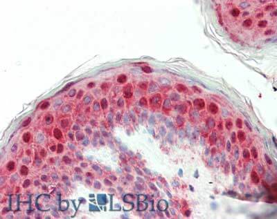
Immunohistochemistry of rabbit anti-Laminin antibody. Tissue: human skin. Fixation: formalin fixed paraffin embedded. Antigen retrieval: not required. Primary antibody: Anti-Laminin at 5 μg/mL for 1 h at RT. Secondary antibody: Peroxidase rabbit secondary antibody at 1:10,000 for 45 min at RT. Staining: Laminin as precipitated red signal with hematoxylin purple nuclear counterstain.
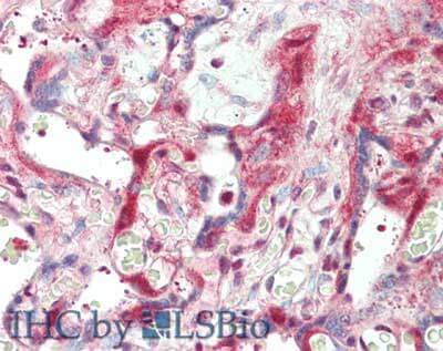
Immunohistochemistry of rabbit anti-Laminin antibody. Tissue: human placenta. Fixation: formalin fixed paraffin embedded. Antigen retrieval: not required. Primary antibody: Anti-Laminin at 5 μg/mL for 1 h at RT. Secondary antibody: Peroxidase rabbit secondary antibody at 1:10,000 for 45 min at RT. Staining: Laminin as precipitated red signal with hematoxylin purple nuclear counterstain.
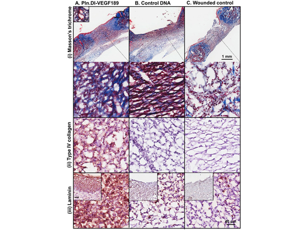
Expression of collagen and keratin (i), collagen type IV (ii) and laminin (iii) in normal rats after 14 days treatmentwith (A) Pln.D1-VEGF189 plasmid DNA, (B) control plasmid DNA loaded chitosan scaffolds or (C) control wounds. Collagen and keratin were detected in panel (i) using Masson's trichrome that stains collagen blue, keratin red and nuclei red/brown. The inset in panel A (i) is a representative blood vessel. The low magnification images in panel A reveal the wound site while the higher magnification images in panels A?C represent the healing region of the wounds. The inset in panels A?C (iii) represent the epidermis. Scale bar represented 1 mm (panel A) and higher magnification images contained a scale bar that represented 40 μm and inset (panel A i and iii). Fig 6. PMID:?28189628
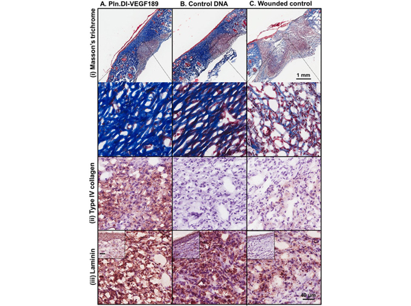
Expression of collagen and keratin (i), collagen type IV (ii) and laminin (iii) in diabetic rats after 28 days of treatment with (A) Pln.D1-VEGF189 plasmid DNA, (B) control plasmid DNA loaded chitosan scaffolds or (C) control wounds. Collagen and keratin were detected in panel (i) using Masson's trichrome that stains collagen blue, keratin red, nuclei and erythrocytes red/brown. The low magnification images in panel A reveal the wound site while the higher magnification images in panels A?C represent the healing region of the wounds. The inset in panels A?C (iii) represent the epidermis. Scale bar represented 1 mm (panel A) and higher magnification images contained a scale bar that represented 40 μm and inset (panels A?C (iii)). Fig 10. PMID:?28189628
|
|
|
|
Immunohistochemistry of rabbit anti-Laminin antibody. Tissue: human skin. Fixation: formalin fixed paraffin embedded. Antigen retrieval: not required. Primary antibody: Anti-Laminin at 5 μg/mL for 1 h at RT. Secondary antibody: Peroxidase rabbit secondary antibody at 1:10,000 for 45 min at RT. Staining: Laminin as precipitated red signal with hematoxylin purple nuclear counterstain.
|
|
| 別品名 |
rabbit anti-Laminin antibody, LAMA1 antibody, LAMB1 antibody, Laminin A chain antibody, Laminin alpha 1 antibody, Laminin alpha 1 chain antibody, Laminin B1 chain antibody, Laminin subunit beta 1 antibody
|
| 別包装 |
あり
|
| 交差種 |
Human
|
| 適用 |
Immunohistochemistry
|
| 免疫動物 |
Rabbit
|
| 標識物 |
Unlabeled
|
| 精製度 |
Affinity Purified
|
| GENE ID |
3912
|
| Accession No.(Gene/Protein) |
AAA59486.1, P07942, P25391
|
| Gene Symbol |
LAMA1/LAMB1
|
| 参考文献 |
[[Cosmo Reference Collection]][Pub Med ID]24517414,28189628
|
| [注意事項] |
濃度はロットによって異なる可能性があります。メーカーDS及びCoAからご確認ください。
|
|
| メーカー |
品番 |
包装 |
|
RKL
|
600-401-116-0.1
|
0.1 MG
|
※表示価格について
| 当社在庫 |
なし
|
| 納期目安 |
約10日
|
| 保存温度 |
4℃
|
|
※当社では商品情報の適切な管理に努めておりますが、表示される法規制情報は最新でない可能性があります。
また法規制情報の表示が無いものは、必ずしも法規制に非該当であることを示すものではありません。
商品のお届け前に最新の製品法規制情報をお求めの際はこちらへお問い合わせください。
|
※当社取り扱いの試薬・機器製品および受託サービス・創薬支援サービス(納品物、解析データ等)は、研究用としてのみ販売しております。
人や動物の医療用・臨床診断用・食品用としては、使用しないように、十分ご注意ください。
法規制欄に体外診断用医薬品と記載のものは除きます。
|
|
※リンク先での文献等のダウンロードに際しましては、掲載元の規約遵守をお願いします。
|
|
※CAS Registry Numbers have not been verified by CAS and may be inaccurate.
|


