|
※サムネイル画像をクリックすると拡大画像が表示されます。
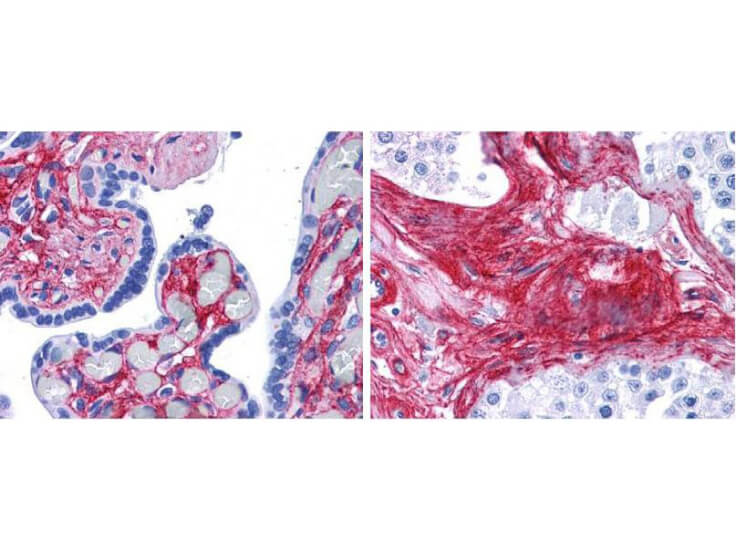
Rockland anti collagen VI antibody (600-401-108 Lot 26009, 1:400 45 min RT) showed strong staining in FFPE sections of human placenta (Left) with red staining of stromal and extracellular spaces, and in testis (Right) with staining of extracellular spaces between seminiferous tubules). Slides were steamed in 0.01 M sodium citrate buffer, pH 6.0 at 99-100°C - 20 minutes for antigen retrieval. Images provided courtesy of LifeSpan Biosciences, Seattle, WA
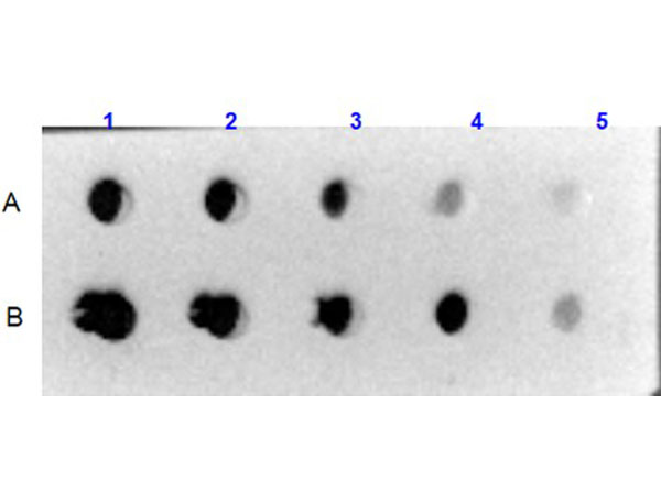
Dot Blot results using Rabbit Anti-Collagen VI Antibody. Sample: A) Human Collagen VI (p/n 009-001-108), B) Bovine/Human Collagen VI (p/n 001-001-108).Load: 1) 1000ng, 2) 333.33ng, 3) 111.11ng, 4) 37.03ng, 5) 12.35ng.Primary Antibody: Rabbit Anti-Collagen VI antibody (p/n 600-401-108) at 5μg/mL at RT for 1hr. Secondary Antibody: Goat Anti-Rabbit IgG HRP Conjugated (p/n 611-1302) 1:40,000 at RT for 30mins. Blocking: BlockOut(R) Universal Blocking Buffer (p/n MB-073) at RT for 30mins.
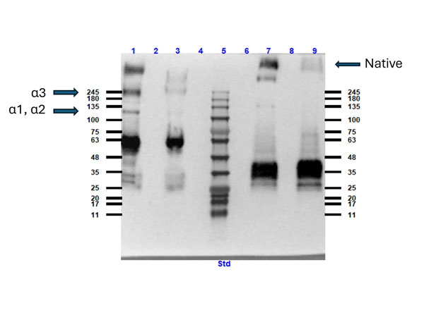
Western blot results using Rabbit Anti-Collagen Type VI Antibody. Lane: 1) Human Collagen Type VI (p/n 009-001-108) reduced and boiled, 3) Human Collagen Type VI reduced and not boiled,5) Opal prestain molecular weight marker (p/n MB-210-0500), 7) Human Collagen Type VI not reduced and boiled, 9) Human Collagen Type VI not reduced and not boiled. Lanes 2, 4, 6, 8 blank. Load: 10μg. Primary Antibody: Rabbit Anti-Collagen Type VI Antibody (p/n 600-401-108) 1:500 overnight at 2-8°C. Secondary Antibody: Goat anti-Rabbit IgG HRP conjugated (p/n 611-1302) 1:40,000 at RT for 30mins. Blocking: BlockOut(R) Universal Blocking Buffer (p/n MB-073) at RT for 1hr.
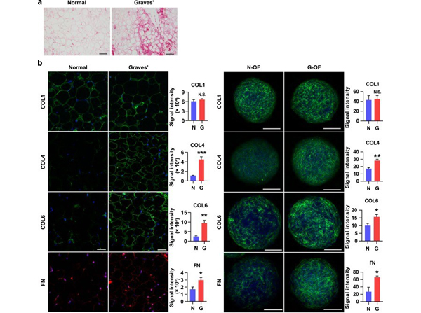
Unique ECM deposition in GO tissues and GO-derived OF organoids. (a) Representative Sirius red staining of periocular adipose tissues in control (normal) and GO (Graves). Scale bars, 100 μm. (b) Representative immunofluorescent staining of collagens (green) and FN (red or green) with DAPI (blue) in control and Graves orbital adipose tissues (left, n = 4 to 5) and 3D organoids reconstituted from hOFs isolated from respective tissues (right). Signal intensities were quantified per spheroid. n = 5 to 8 organoids. Scale bars, 50 μm (left) and 100 μm (right). G indicates G-OF; N indicates N-OF. *P?< 0.05, unpaired Student?t?test (n = 4 in each group). Fig 2. PMID:?30388216
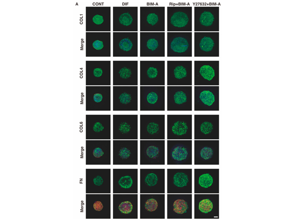
Representative confocal images showing the expression of ECMs in 3D 3T3-L1 spheroids under several sets of conditions. (A) On Day 7, the 3D cultures of spheroids of 3T3-L1 preadipocytes as the control (CONT), and their adipogenic differentiation in the absence (DIF) or presence of 100 nM bimatoprost free acid (BIM-A) and/or 10 μM ROCK-i (Rip or Y27632), were immunostained with specific antibodies of ECMs designated by the green color. Scale bar: 100 μm. Fig 9.PMID:?36421103
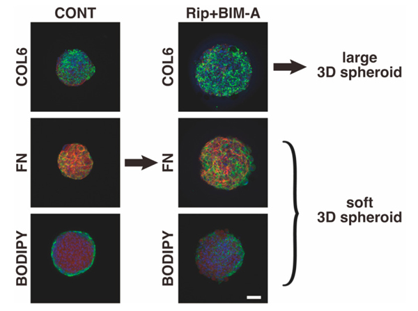
Hypothetical model of the mechanism responsible for inducing huge-sized and soft 3D spheroids caused by the addition of Rip to BIM-A. Fig 10. PMID:?36421103
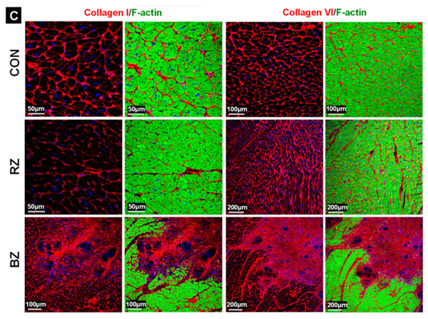
(C) Staining of collagen I and VI mark fibrotic areas. For comparison, the control (CON) tissue is shown. Border zone (BZ) is the myocardial area surrounding the granuloma and remote zone (RZ) is the myocardium distant to granuloma. The nuclei are stained blue with DAPI.?Fig 1. PMID:?33923774
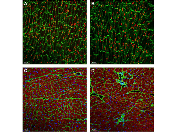
Representative vinculin staining (green) in WT (PBS) mice (A) and WT doxorubicin (B). Nuclei (red) were visualized with draq5. Replacement fibrosis was detected in the doxorubicin treated hearts (D) but not in vehicle hearts (C), visualising with the anti-collagen VI antibody (green). Nuclei (blue) were visualised with DAPI, (E, F). Fig 2. PMID:?28662206
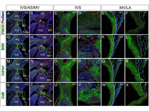
Immunolocalization of FMOD (green, A-F), BGN (green, G-L), Col1a1 (green, M-R), and Col6 (green, S-X) in?Fmod+/+?(A, C, E, G, I, K, M, O, Q, S, U, W) and?Fmod-/-?(B, D, F, H, J, L, N, P, R, T, V, X) mitral leaflet attachments. White boxes in A, B, G, H, M, N, S, T are magnified in panels C, D, I, J, O, P, U, V respectively. Yellow boxes in A, B, G, H, M, N, S, T are magnified and shown in E, F, K, L, Q, R, W, X respectively. Red- propidium iodide (PI); blue- α-sarcomeric actin (αSarc); IVS-interventricular septum; AS- atrial septum; MV- mitral valve; LA- left atrium; solid lines- distinct separation of MV and LA; dashed lines- diffuse attachment of left atrial to MV. Scale bars: A= 400μm applies to B, G, H, M, N, S, T; C= 50μm applies to D-F, I-L, O-R, U-X. Fig 3. PMID:?27503167
|
|
|
|
Rockland anti collagen VI antibody (600-401-108 Lot 26009, 1:400 45 min RT) showed strong staining in FFPE sections of human placenta (Left) with red staining of stromal and extracellular spaces, and in testis (Right) with staining of extracellular spaces between seminiferous tubules). Slides were steamed in 0.01 M sodium citrate buffer, pH 6.0 at 99-100°C - 20 minutes for antigen retrieval. Images provided courtesy of LifeSpan Biosciences, Seattle, WA
|
|
| 別品名 |
rabbit anti-Collagen Type VI antibody, Collagen alpha-1 (VI) chain, Collagen VI antibody, Human mRNA for collagen VI alpha 1 C terminal globular domain antibody
|
| 別包装 |
あり
|
| 由来詳細 |
[Species]Human/Bovine
|
| 交差種 |
Human
Bovine
|
| 適用 |
Immunohistochemistry
Dot Blot
|
| 免疫動物 |
Rabbit
|
| 標識物 |
Unlabeled
|
| 精製度 |
Affinity Purified
|
| GENE ID |
1291
|
| Accession No.(Gene/Protein) |
AAH52575.1, P12109, P12110, P12111
|
| Gene Symbol |
COL6A1-COL6A3
|
| 参考文献 |
[[Cosmo Reference Collection]][Pub Med ID]32409724,24529431,29540474,30388216,17672847,27377666,27503167,28662206,35523828,35329980,33923774,36421103,37878345
|
| [注意事項] |
濃度はロットによって異なる可能性があります。メーカーDS及びCoAからご確認ください。
|
|
| メーカー |
品番 |
包装 |
|
RKL
|
600-401-108-0.1
|
0.1 MG
|
※表示価格について
| 当社在庫 |
なし
|
| 納期目安 |
約10日
|
| 保存温度 |
4℃
|
|


