|
※サムネイル画像をクリックすると拡大画像が表示されます。
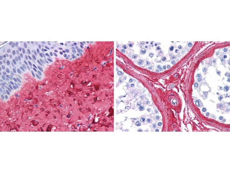
Rockland anti collagen III antibody (600-401-105 Lot 26016, 1:400, 45 min RT) showed strong staining in FFPE sections of human skin(left, dermis) with moderate to strong red staining and testis (right) where strong staining was observed within connective tissue between seminiferous tubules. The antibody showed strong extracellular staining within connective tissues across many organs with minimal background staining. Slides were steamed in 0.01 M sodium citrate buffer, pH 6.0 at 99-100°C - 20 minutes for antigen retrieval. Images provided courtesy of LifeSpan Biosciences, Seattle, WA
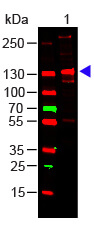
Western Blot of Rabbit Anti-COLLAGEN III Antibody. Lane 1: Human Collagen III (p/n 009-001-105). Load: 100ng per lane. Primary antibody: Collagen III Antibody at 1:1000 o/n at 4°C. Secondary antibody: DyLight? 649 Goat anti-rabbit (p/n 611-1302) at 1:20,000 for 30 min at RT. Block: MB-070 for 30 min at RT. Predicted/Observed size: 138 kDa, 138 kDa.
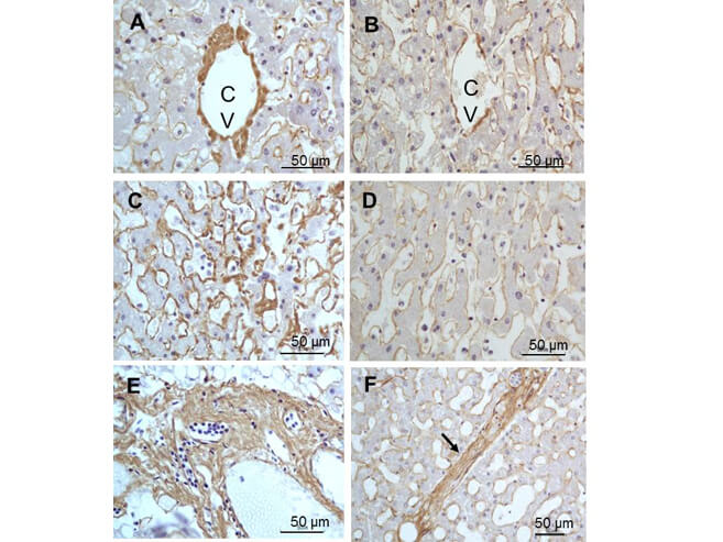
Immunohistochemistry of Rabbit Anti-collagen type III antibody. Tissue: right lobe of the liver section. A:Central Vein (CV) fibrosis, B: Non-fibrotic CV, C: Perisinusodial fibrosis, D: Non-fibrotic area, E: Protat tract fibrosis, F: Septal fibrosis (arrow). Fixation: formalin fixed paraffin embedded. Antigen retrieval: not required. Primary antibody: Anti-collagen type III at 1:500 for 4°C for 24hr. Secondary antibody: Peroxidase biotin-streptavidin rabbit secondary antibody at 1:10,000 for 45 min at RT. Localization: Anti-collagen type III is intra and extracellular. Staining: 3.3’-diaminobenzidine tetrahydrochloride was used as the chromogen. Nuclei were counterstained purple with hematoxylin.
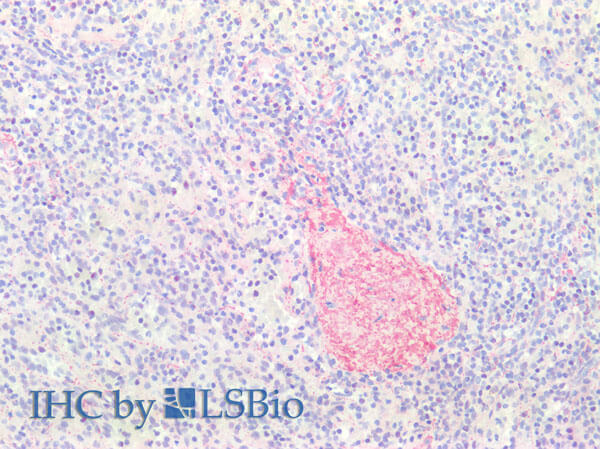
Immunohistochemistry of Rabbit Anti-Collagen Type III Antibody. Tissue: FFPE normal human spleen tissues (10X). Antigen Retrieval: 0.01 M sodium citrate buffer for 20 minutes. Primary Antibody: Anti-Collagen Type III at 5μL/mL for 45 mins at RT. Staining: Anti-Rabbit biotinylated secondary antibody for 30 min at RT. Alkaline phosphatase streptavidin for 30 min at RT. Alkaline phosphatase chromogen substrate for 30 min at RT. The stained slides were evaluated by a pathologist to confirm staining specificity.
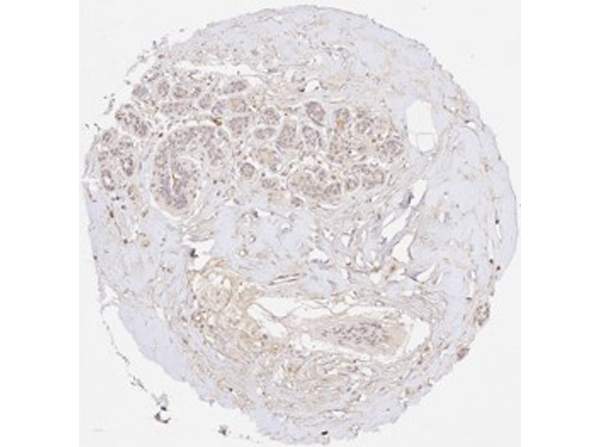
Immunohistochemistry results of Rabbit Anti-Collagen Type I Antibody. Tissue: human stroma in colorectal cancer. Fixation: FFPE. Antigen Retrieval: HIER using Tris-EDTA-citrate buffer pH 7.8 for 5 min. Blocking: Peroxidase-Blocking Solution for 10 min. Primary Antibody: Anti-Collagen Type I (p/n 600-401-103-0.1) at 1:15 for 1 hr at 37 °C. Secondary Antibody: Dako REAL EnVision Detection Kit, Polymer-HRP, Rabbit/Mouse. Counterstain: Hematoxylin for 15 sec. Substrate: DAB-Chromogen, Rabbit/Mouse. Staining/Results: Fibrillar collagen III staining of the stroma in a colorectal cancer. Independently Validated by antibodies-online GmbH (p/n ABIN7565873/ ABIN5596830/ ABIN5596829) courtesy of MS Validated Antibodies.
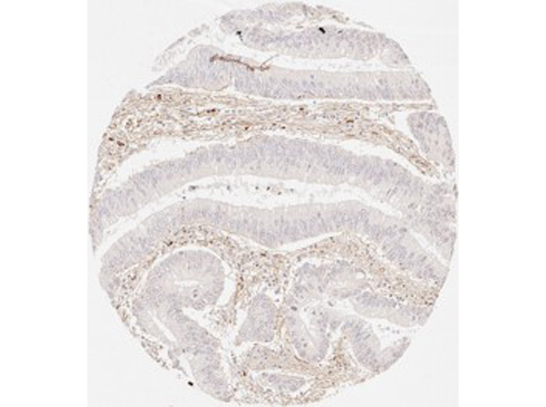
Immunohistochemistry results of Rabbit Anti-Collagen Type III Antibody. Tissue: human oral cavity. Fixation: FFPE. Antigen Retrieval: HIER using Tris-EDTA-citrate buffer pH 7.8 for 5 min. Blocking: Peroxidase-Blocking Solution for 10 min. Primary Antibody: Anti-Collagen Type III (p/n 600-401-105-0.1) at 1:15 for 1 hr at 37 °C. Secondary Antibody: Dako REAL EnVision Detection Kit, Polymer-HRP, Rabbit/Mouse. Counterstain: Hematoxylin for 15 sec. Substrate: DAB-Chromogen, Rabbit/Mouse. Staining/Results: Distinct fibrillar collagen III staining of the stroma in squamous cell carcinoma of the oral cavity. Independently Validated by antibodies-online GmbH (p/n ABIN7565873/ ABIN5596830/ ABIN5596829) courtesy of MS Validated Antibodies.
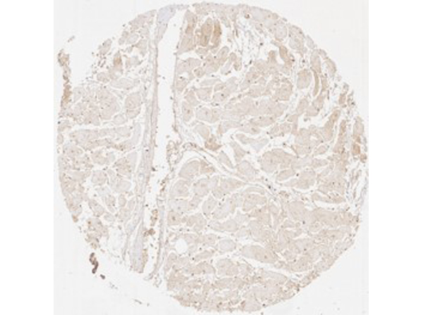
Immunohistochemistry results of Rabbit Anti-Collagen Type III Antibody. Tissue: human heart muscle. Fixation: FFPE. Antigen Retrieval: HIER using Tris-EDTA-citrate buffer pH 7.8 for 5 min. Blocking: Peroxidase-Blocking Solution for 10 min. Primary Antibody: Anti-Collagen Type III (p/n 600-401-105-0.1) at 1:15 for 1 hr at 37 °C. Secondary Antibody: Dako REAL EnVision Detection Kit, Polymer-HRP, Rabbit/Mouse. Counterstain: Hematoxylin for 15 sec. Substrate: DAB-Chromogen, Rabbit/Mouse. Staining/Results: Distinct fibrillar collagen III staining surrounding each heart muscle cell. Independently Validated by antibodies-online GmbH (p/n ABIN7565873/ABIN5596830/ABIN5596829) courtesy of MS Validated Antibodies.
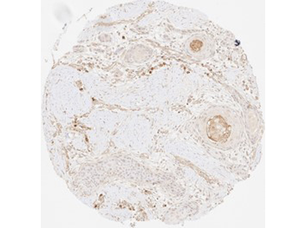
Immunohistochemistry results of Rabbit Anti-Collagen Type III Antibody. Tissue: human non-cancerous breast tissue. Fixation: FFPE. Antigen Retrieval: HIER using Tris-EDTA-citrate buffer pH 7.8 for 5 min. Blocking: Peroxidase-Blocking Solution for 10 min. Primary Antibody: Anti-Collagen Type III (p/n 600-401-105-0.1) at 1:225 for 1 hr at 37 °C. Secondary Antibody: Dako REAL EnVision Detection Kit, Polymer-HRP, Rabbit/Mouse. Counterstain: Hematoxylin for 15 sec. Substrate: DAB-Chromogen, Rabbit/Mouse. Staining/Results: Fibrillar collagen III staining in non-cancerous breast tissue showing considerable sclerosis. Independently Validated by antibodies-online GmbH (p/n ABIN7565873/ ABIN5596830/ ABIN5596829) courtesy of MS Validated Antibodies.
(a) Typical electropherogram of α (I) subunits, oligomers β/γ (I), and collagen type I degradation products, released from healthy and burnt skin. Collagenous components released from tissue samples using pepsin were submitted to 4?15% gradient SDS-PAGE, in nonreducing conditions. (b) Interferences of collagen type III, in electrophoretic profiles of collagen type I components extracted from healthy and burned skin. Collagen components were submitted to electrophoresis in the absence of dithiothreitol (reducing disulfide bonds) and subsequently―after electrotransfer to Immobilon―subjected to reaction with collagen type III antibodies. Lane 1: components of collagen type I isolated from healthy skin. Lane 2: components of collagen type I isolated from burned skin treated with propolis. Lane 3: components of collagen type I isolated from burned skin treated with AgSD. Lane 4: components of collagen type I isolated from burned skin treated with propolis vehicle. Lane 5: components of collagen type I isolated from burned skin treated with NaCl. Figure provided by CiteAb. Source: Evid Based Complement Alternat Med, PMID: 23781260.
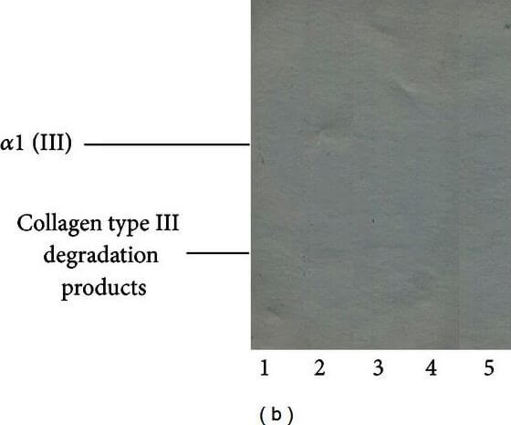
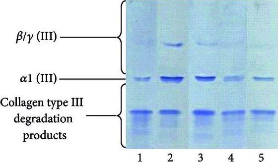
Typical electropherogram of α (III) subunits, oligomers β/γ (III), and collagen type III degradation products, released from healthy and burned skin. Collagenous components released from tissue samples using pepsin were submitted to 4?15% gradient SDS-PAGE, western blotted, and probed with collagen type III antibodies. Lane 1: components of collagen type I isolated from healthy skin. Lane 2: components of collagen type I isolated from burned skin treated with propolis. Lane 3: components of collagen type I isolated from burned skin treated with AgSD. Lane 4: components of collagen type I isolated from burned skin treated with propolis vehicle. Lane 5: components of collagen type I isolated from burned skin treated with NaCl. Figure provided by CiteAb. Source: Evid Based Complement Alternat Med, PMID: 23781260.
|
|
|
|
Rockland anti collagen III antibody (600-401-105 Lot 26016, 1:400, 45 min RT) showed strong staining in FFPE sections of human skin(left, dermis) with moderate to strong red staining and testis (right) where strong staining was observed within connective tissue between seminiferous tubules. The antibody showed strong extracellular staining within connective tissues across many organs with minimal background staining. Slides were steamed in 0.01 M sodium citrate buffer, pH 6.0 at 99-100°C - 20 minutes for antigen retrieval. Images provided courtesy of LifeSpan Biosciences, Seattle, WA
|
|
| 別品名 |
rabbit anti-Collagen Type III antibody, Collagen type III alpha 1 antibody, Collagen type III alpha antibody, EDS4A antibody, Ehlers Danlos syndrome type IV, autosomal dominant antibody, Fetal collagen antibody, COL3A1, Collagen alpha-1 (III) chain
|
| 由来詳細 |
[Species]Human/Bovine
|
| 交差種 |
Human
Bovine
Porcine
|
| 適用 |
Western Blot
Immunohistochemistry
Dot Blot
|
| 免疫動物 |
Rabbit
|
| 標識物 |
Unlabeled
|
| 精製度 |
Affinity Purified
|
| GENE ID |
1281
|
| Accession No.(Gene/Protein) |
NP_000081.1, P02461
|
| Gene Symbol |
COL3A1
|
| 参考文献 |
[Pub Med ID]32409493
|
| [注意事項] |
濃度はロットによって異なる可能性があります。メーカーDS及びCoAからご確認ください。
|
|
| メーカー |
品番 |
包装 |
|
RKL
|
600-401-105-0.5
|
0.5 MG
|
※表示価格について
| 当社在庫 |
なし
|
| 納期目安 |
約10日
|
| 保存温度 |
4℃
|
|
