|
※サムネイル画像をクリックすると拡大画像が表示されます。
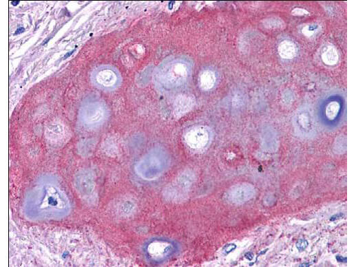
Rockland anti collagen II antibody (600-401-104 Lot 26014, 1:400, 45 min RT) showed moderate staining of in FFPE human bronchiolar cartilage (shown in image). Though not shown, faint to moderate staining of tonsillar squamous epithelium, prostatic stroma, breast, colon, placenta, and dermal connective tissues was also observed. All other tissues, including brain, breast epithelium, colon epithelium, heart, intestine, kidney, liver, lung, skeletal muscle, pancreas, spleen, testis, thymus, thyroid, and uterus were negative for staining Slides were steamed in 0.01 M sodium citrate buffer, pH 6.0 at 99-100°C - 20 minutes for antigen retrieval. Image provided courtesy of LifeSpan Biosciences, Seattle, WA
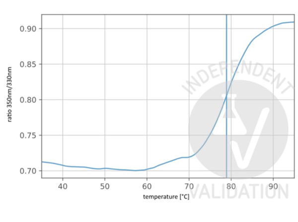
Unfolding profile of Anti-Collagen Type II (RABBIT) Antibody (p/n 600-401-104). The fluorescence signal is plotted against temperature. The vertical line indicates the Ti at 78.9°C. Independently Validated by?antibodies-online GmbH (p/n ABIN7565872/ ABIN5596824/ ABIN5596825) courtesy of?NanoTemper Technologies.
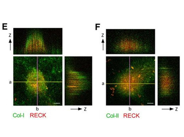
(E and F) Localization of type I and type II collagen at cartilaginous nodules formed by ATDC5 cells. ATDC5 cells incubated for 15 days were fixed and stained with anti-RECK (red) plus anti-type I collagen (green; E) or anti-RECK (red) plus anti-type II collagen (green; F). Reconstituted z-axis images along two cutting lines (a,b) are shown in the top and right panels, respectively. Bars, 100 μm in E,F. Fig 2. PMID:?17298979
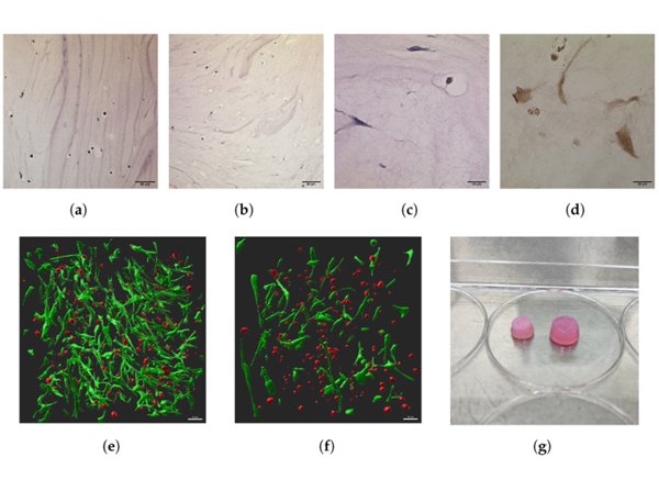
(a?d) Show photos of histological sections. (a,b) HE stainings of D4 at 10×: (a) is the control, (b) is the ES group. (c) An HE staining of the D7 ES group at 40×; (d) a COL2 immunohistochemical staining from the D10 ES group at 40× magnification. (e,f) Show 3D renderings of stacks from two-photon imaging from D7. (e) Displays the ES group; (f) the control group. (g) Shows a physical image of the scaffolds at D10. The left scaffold is from the ES group, the right scaffold is from the control group. Fig 3. PMID:?38927763
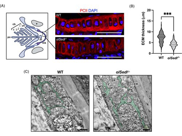
Col2 expression and secretion is compromised in?olSedl-/-?fish. (A) Representative immunostaining of Meckel's cartilage (boxed area in the graphic representation on the left) of WT and?olSedl-/-?fish showing a reduction in type II collagen (red) levels. Nuclei are counterstained with DAPI (blue) [(cb1-4) ceratobranchial pairs 1 to 4, (ch) ceratohyal, (et) ethmoid plate, (mk) Meckel's cartilage, (po) posterior limit, (e) eye]. (B) Quantification of ECM thickness in WT and?olSedl-/-?fish. (C) Representative electron microscopy images (16× magnification) of a vertebral section of stage 40 WT and?olSedl-/-?fish. The ER is pseudocolored in green and the ER cisternae are indicated by black arrows. The dashed line and punctate pattern in the violin plot in (B) show the median and quartiles, respectively. (B):?n?>?90 measurements. ***p?<?0.001, two-tailed unpaired?t-test with Welch's correction. Scale bar in (A): 30?μm, (C): 500?nm. Fig 2. PMID:?37963679
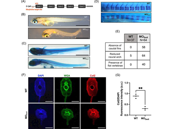
olSedl?morpholino fish manifest a SEDT-like phenotype. (A) Schematic representation of the morpholino targeting site at the ATG in exon 1 of?olSedl. (B) Representative brightfield images of WT and MOSedl?fish. (C) Representative whole-mount alcian blue-alizarin red staining images showing that MOSedl?lack cartilaginous intermediates in the caudal fin and have a reduced number of vertebrae. (D) High magnification alcian blue-alizarin red staining from the first to tenth vertebra showing altered morphology. (E) Quantification of the observed phenotypes. (F) Immunofluorescence analysis of WT and MOSedl?sections of frontal vertebra showing a significant alteration in size and morphology. Nuclei are counterstained with DAPI (blue), connective tissue is visualized with fluorescent Wheat Germ Agglutinin (WGA, green), while type II collagen (Col2) is shown in red. (G) Col2 fluorescence intensity measurement of WT and MOSedl?sections of frontal vertebra;?N?=?3, median is indicated. **p?=?0.001, two-tailed unpaired?t-test. Scale bar in (B) and (C): 1?mm; (F): 100?μm. Fig 3. PMID:?37963679
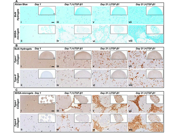
Histological characterisation of bulk hydrogels and NHSA-microgels at day 1, 7 & 21 of?in vitro?culture. (A) Alcian blue (pH 1) staining ? bulk hydrogel (top row) & NHSA-microgels (bottom row). (B) Type-I and type-II collagen immunohistochemical staining on bulk hydrogels. (C) Type-I and type-II collagen immunohistochemical staining on NHSA-microgels (scale bar: 50 μm/inset: 500 μm). Fig 5. PMID:?31994552
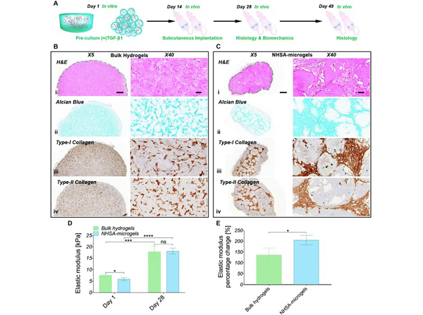
(A) Schematic representation of hCC-laden bulk hydrogels and NHSA-microgel?in vivo?chondrogenesis: after?in vitro?pre-culture in chondrogenic media ((+)TGF-β1) for two weeks, samples were subcutaneously implanted into nude mice (left mice ? bulk hydrogel; right mice ? NHSA-microgels) and maintained?in vivo?for an additional two weeks (day 28) or five weeks (day 49). (B) Histological analysis of bulk hydrogel at day 28 (i: H&E, ii: Alcian blue, iii: type-I collagen, iv: type-II collagen). (C) Histological analysis of NHSA-microgel at day 28 (i: H&E, ii: Alcian blue, iii: type-I collagen, iv: type-II collagen) (scale bar: X5/500 μm; X40/50 μm). (D) Nanoindentation tests at day 1 and day 28. (E) Percentage elastic modulus increment profile showing hCC-laden bulk hydrogel and NHSA-microgel mechanical properties enhancement after 28 days maintenance?in vivo?(*P?< 0.05, **P?< 0.01, ***P?< 0.001, and ****P?< 0.0001). Fig 7. PMID:?31994552
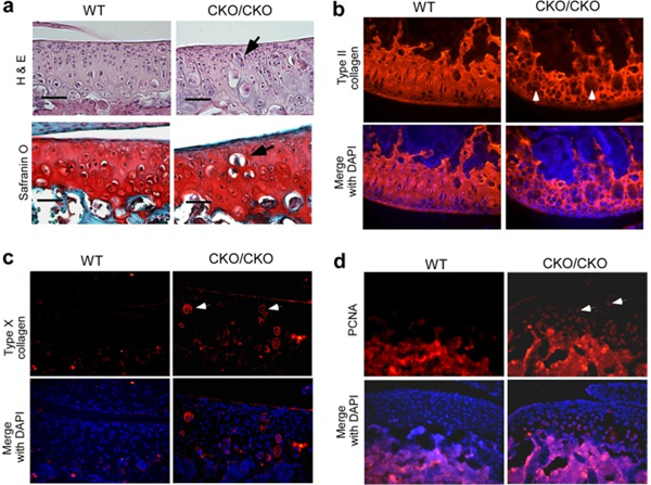
Articular chondrocyte hypertrophy and proliferation in 1-month-old ESET-null mice.?a, 1-month-old knee joints were stained by H&E and Safranin O for detailed morphological analysis.?b, 1-month-old knee sections were stained with an anti-type II collagen antibody. The type II collagen network was stained?red, and nuclei were stained?blue?by DAPI.?c, anti-type X collagen antibody specifically stains hypertrophic chondrocytes (red) in 1-month-old knee sections.?d, anti-PCNA antibody detects cells positive for the proliferation marker in 1-month-old knee sections.?Arrows?in?a?d?indicate locations of hypertrophic or proliferative chondrocytes near the superficial zone in ESET-null mice. A total of three wild-type and five knock-out mice were examined, and images from representative experiments are shown. Genotypes of the mice are indicated on the?top. Scale bar: 50 μm. Fig 3. PMID:?24056368
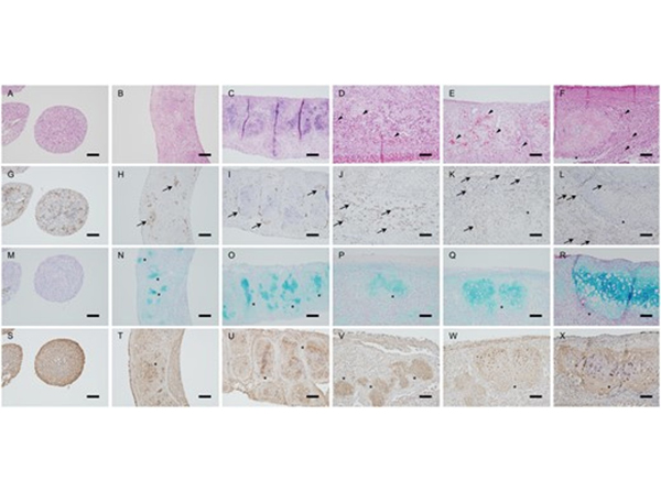
Histological findings of the spheroids, artificial tracheas and transplanted grafts. (A?F) Haematoxylin and eosin staining, (G?L) immunohistochemical staining with anti-CD31 antibodies, (M?R) Alcian blue staining and (S?X) immunohistochemical staining with anti-collagen II antibodies. (A,?G,?M?and?S) Spheroids; (B,?H,?N?and?T) bio-3D-printed artificial tracheas after maturation; (C,?I,?O?and?U) Day 1; (D,?J,?P?and?V) Day 8; (E,?K,?Q?and?W) Day 11 and (F,?L,?R?and?X) Day 23. Cartilage tissue can be observed in artificial tracheas after maturation and maintained over 10?days (asterisk). Capillary-like tubes consisting of CD31-positive cells (arrow) can be seen in artificial tracheas after maturation. Red blood cells are observed from Day 8 in the capillary-like tube formations (arrowhead). Scale bar?=?100μm. Fig 5. PMID:?29346562
|
|
|
|
Rockland anti collagen II antibody (600-401-104 Lot 26014, 1:400, 45 min RT) showed moderate staining of in FFPE human bronchiolar cartilage (shown in image). Though not shown, faint to moderate staining of tonsillar squamous epithelium, prostatic stroma, breast, colon, placenta, and dermal connective tissues was also observed. All other tissues, including brain, breast epithelium, colon epithelium, heart, intestine, kidney, liver, lung, skeletal muscle, pancreas, spleen, testis, thymus, thyroid, and uterus were negative for staining Slides were steamed in 0.01 M sodium citrate buffer, pH 6.0 at 99-100°C - 20 minutes for antigen retrieval. Image provided courtesy of LifeSpan Biosciences, Seattle, WA
|
|
| 別品名 |
rabbit anti-Collagen Type II antibody, Collagen alpha-1 (II) chain, Alpha-1 type II collagen, Cartilage collagen antibody, Chondrocalcin antibody, COL2A1 antibody
|
| 別包装 |
あり
|
| 由来詳細 |
[Species]Human/Bovine
|
| 交差種 |
Human
Bovine
|
| 適用 |
Immunohistochemistry
Dot Blot
|
| 免疫動物 |
Rabbit
|
| 標識物 |
Unlabeled
|
| 精製度 |
Affinity Purified
|
| GENE ID |
1280
|
| Accession No.(Gene/Protein) |
NP_001835.3, P02458
|
| Gene Symbol |
COL2A1
|
| 参考文献 |
[[Cosmo Reference Collection]][Pub Med ID]38927763,28084417,17298979,30559329,17785842,19660048,23107560,24056368,25160583,29624608,30862915,31534979,31536840,31932796,32078578,32223949,27885831,28273419,32011382,32578533+他多数
|
| [注意事項] |
濃度はロットによって異なる可能性があります。メーカーDS及びCoAからご確認ください。
|
|
| メーカー |
品番 |
包装 |
|
RKL
|
600-401-104-0.5
|
0.5 MG
|
※表示価格について
| 当社在庫 |
なし
|
| 納期目安 |
約10日
|
| 保存温度 |
4℃
|
|


