|
※サムネイル画像をクリックすると拡大画像が表示されます。
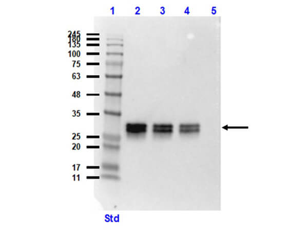
Western blot of Mouse Anti-GFP Antibody. Lane 1: Opal Prestained Molecular Weight Marker (p/n MB-210-0500). Lane 2: HeLa WC Lysate+GFP protein (p/n W09-000-364 [10μg]/ p/n 000-001-215 [50ng]). Lane 3: HeLa WC Lysate+GFP protein (10μg/20ng). Lane 4: HeLa WC Lysate+GFP protein (10μg/10ng). Lane 5: HeLa Whole Cell Lysate (p/n W09-000-364) (10μg). Primary Antibody: Anti-GFP at 1:1000 overnight at 2-8°C. Secondary Antibody: Rabbit Anti-Mouse IgG HRP (p/n 610-4302) at 1:40,000 for 30mins at RT. Block: BlockOut Buffer (p/n MB-073). Expected MW: ~27kDa.
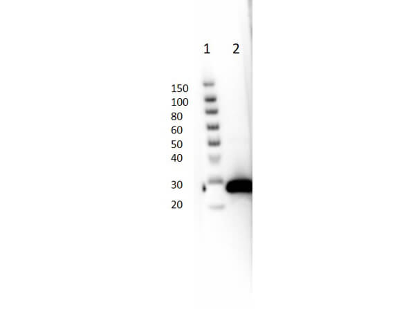
Western blot of Mouse Anti-GFP Antibody. Lane 1: Thermo SuperSignal Molecular Weight Marker. Lane 2: GFP protein (p/n 000-001-215) [50ng]. Primary Antibody: Anti-GFP at 1:1000 overnight at 2-8°C. Secondary Antibody: Rabbit Anti-Mouse IgG HRP (p/n 610-4302) at 1:40,000 for 30mins at RT. Block: BlockOut Buffer (p/n MB-073). Expected MW: ~27kDa.
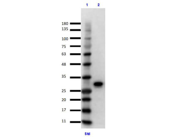
Immunoprecipitation/Western Blot using GFP Protein. Lane 1: Opal Prestained Molecular Weight Marker (p/n MB-210-0500). Lane 2: GFP Input (p/n 000-001-215) Reduced [10μL]. Primary IP Antibody: Mouse Anti-GFP (p/n 600-301-215) at 10μg overnight at 2-8°C. Secondary Antibody: TrueBlot Anti-Mouse Ig IP Agarose Beads (p/n 00-8811-25) at 500μg for 1hr at RT. Buffer: BlockOut Buffer (p/n MB-073) for 30 mins at RT. Exposure: 7 sec.
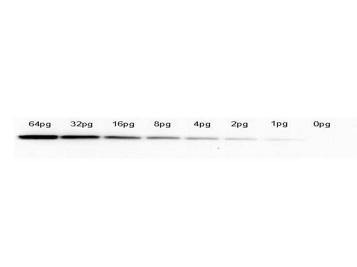
Western Blot of anti-GFP monoclonal antibody. Lane 1: 64pg of recombinant GFP protein (p/n 000-001-215) were spiked into a HeLa cell-derived lysates (p/n W09-000-364). Lane 2: 32pg of recombinant GFP protein were spiked into a HeLa cell-derived lysates. Lane 3: 16pg of recombinant GFP protein were spiked into a HeLa cell-derived lysates. Lane 4: 8pg of recombinant GFP protein were spiked into a HeLa cell-derived lysates. Lane 5: 4pg of recombinant GFP protein were spiked into a HeLa cell-derived lysates. Lane 6: 2pg of recombinant GFP protein were spiked into a HeLa cell-derived lysates. Lane 7: 1g of recombinant GFP protein were spiked into a HeLa cell-derived lysates. Lane 8: 0pg of recombinant GFP protein were spiked into a HeLa cell-derived lysates. Primary antibody: anti-GFP monoclonal antibody at 1:400 for overnight at 4°C. Secondary antibody: HRP-conjugated anti-Mouse IgG (p/n 610-4302) was performed at a dilution of 1:20,000 for 1h at 4°C. Block: TTBS (p/n MB-013) supplemented with 1% BSA (p/n BSA-50) for 1 h at 4°C. Predicted/Observed size: 27 kDa for GFP. Other band(s): none.
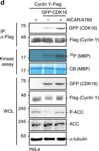
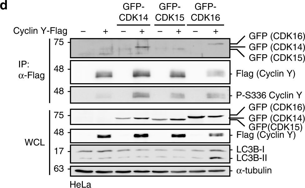
Protein microarray screen for the identification of AMPK substrates.a Schematic representation of the ProtoArray based screen with approximately 9000 human proteins using AMPK (see also Supplementary Data 1). b Details of two sub-arrays incubated with or without AMPK with marked substrates are shown. c GST-CDK16, Cyclin Y-His6 and GST were incubated in the presence of [γ-32P]-ATP with AMPK. Phosphorylation was determined by autoradiography (32P, top). Proteins were visualized by Coomassie blue staining (CB, bottom; n?=?2). d HeLa cells were transfected with vectors expressing GFP-CDK16 and Cyclin Y-Flag and treated for 1?h with 0.5?mM AICAR/50?μM A769662 (A769) as indicated. Cyclin Y-Flag was immunoprecipitated with Flag antibodies (IP) and immunoblotted against CDK16 and Cyclin Y or used for in vitro kinase assays with myeloid basic protein (MBP) as substrate. Autoradiographs (32P) and Coomassie blue staining (CB) of MBP are displayed. Whole cell lysates (WCL) were immunoblotted with the indicated antibodies (n?=?3). e Quantification of CDK16 co-immunoprecipitated with Cyclin Y. Statistical significance was measured via unpaired and two-tailed Student’s t-tests and is presented as follows: **p?<?0.01, and ***p?<?0.001. All error bars indicate SD (n?=?3; Cyclin Y?+?AICAR/A769 vs. Cyclin Y/CDK16: t?=?8.719, df?=?4; Cyclin Y/CDK16 vs. Cyclin Y/CDK16?+?AICAR/A769: t?=?5.595, df?=?4). n biological independent replicate. SD standard deviation. Source data are provided as a Source Data file. Figure provided by CiteAb. Source: Nat Commun, PMID: 32098961.
Active Cyclin Y/CDK16 complexes induce autophagy.a NIH3T3 cells stably expressing mCherry-GFP-LC3 were transfected with HA-CDK16 and Cyclin Y-Flag as indicated or treated for 2?h with EBSS or for 4?h with 200?nM Bafilomycin A1 (Baf. A1) and lysates were immunoblotted as indicated. KR kinase-deficient CDK16 mutant, AA CDK16 binding deficient Cyclin Y mutant (n?=?3). b Representative confocal images of the NIH3T3-mCherry-GFP-LC3 cells treated as in panel a. Staining of the HA-CDK16 in purple identified transfected cells. Autophagosomes (yellow dots) and autolysosomes (red dots) were detected by an overlay of the GFP and mCherry fluorescent signals. Scale bar: 20?μm. c Quantification of autophagosomes (yellow dots) and autolysosomes (red dots) of cells shown in panel b. Statistical significance was measured via unpaired and two-tailed Student’s t-tests and is presented as follows: **p?
|
|
|
|
Western blot of Mouse Anti-GFP Antibody. Lane 1: Opal Prestained Molecular Weight Marker (p/n MB-210-0500). Lane 2: HeLa WC Lysate+GFP protein (p/n W09-000-364 [10μg]/ p/n 000-001-215 [50ng]). Lane 3: HeLa WC Lysate+GFP protein (10μg/20ng). Lane 4: HeLa WC Lysate+GFP protein (10μg/10ng). Lane 5: HeLa Whole Cell Lysate (p/n W09-000-364) (10μg). Primary Antibody: Anti-GFP at 1:1000 overnight at 2-8°C. Secondary Antibody: Rabbit Anti-Mouse IgG HRP (p/n 610-4302) at 1:40,000 for 30mins at RT. Block: BlockOut Buffer (p/n MB-073). Expected MW: ~27kDa.
|
|
| 別品名 |
mouse anti-GFP antibody, Green Fluorescent Protein, GFP antibody, Green Fluorescent Protein antibody, EGFP, enhanced Green Fluorescent Protein, Aequorea victoria, Jellyfish
|
| 適用 |
Western Blot
Enzyme Linked Immunosorbent Assay
Immunoprecipitation
Dot Blot
|
| 免疫動物 |
Mouse
|
| クローン |
9F9.F9
|
| 抗体クラス |
IgG1κ
|
| 標識物 |
Unlabeled
|
| 精製度 |
Affinity Purified
|
| Accession No.(Gene/Protein) |
P42212
|
| Tag情報 |
GFP
|
| 参考文献 |
[Pub Med ID]39205192
|
| [注意事項] |
濃度はロットによって異なる可能性があります。メーカーDS及びCoAからご確認ください。
|
|
| メーカー |
品番 |
包装 |
|
RKL
|
600-301-215
|
1 MG
|
※表示価格について
| 当社在庫 |
なし
|
| 納期目安 |
約10日
|
| 保存温度 |
-20℃
|
|
※当社では商品情報の適切な管理に努めておりますが、表示される法規制情報は最新でない可能性があります。
また法規制情報の表示が無いものは、必ずしも法規制に非該当であることを示すものではありません。
商品のお届け前に最新の製品法規制情報をお求めの際はこちらへお問い合わせください。
|
※当社取り扱いの試薬・機器製品および受託サービス・創薬支援サービス(納品物、解析データ等)は、研究用としてのみ販売しております。
人や動物の医療用・臨床診断用・食品用としては、使用しないように、十分ご注意ください。
法規制欄に体外診断用医薬品と記載のものは除きます。
|
|
※リンク先での文献等のダウンロードに際しましては、掲載元の規約遵守をお願いします。
|
|
※CAS Registry Numbers have not been verified by CAS and may be inaccurate.
|
