|
※サムネイル画像をクリックすると拡大画像が表示されます。
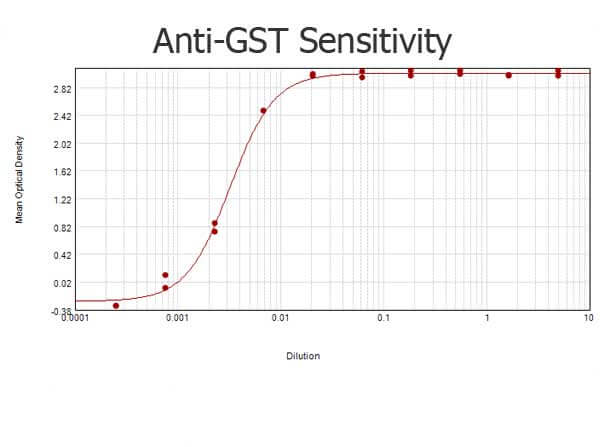
ELISA results of purified Goat anti-GST Antibody Biotin Conjugated tested against purified GST. Each well was coated in duplicate with 1.0 μg of GST (p/n 000-001-200). The starting dilution of antibody was 5μg/ml and the X-axis represents the Log10 of a 3-fold dilution. This titration is a 4-parameter curve fit where the IC50 is defined as the titer of the antibody. Assay performed using 3% fish gelatin as blocking buffer, Streptavidin Peroxidase Conjugated and TMB substrate p/n TMBE-1000
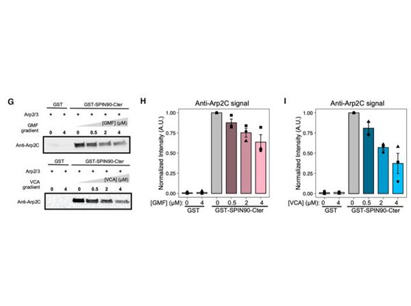
(G?I) GMF and VCA interfere with the SPIN90‐Arp2/3 interaction in the absence of filament. (G) Immunoblots from pull‐down assays where GST beads, decorated with GST or GST‐SPIN90‐Cter (the functional domain of SPIN90), were incubated with 0.4 μΜ Arp2/3 and gradient concentrations of GMF or VCA for 1 h at room temperature. After the unbound protein was washed out, the amount of Arp2/3 attached to the beads was detected by anti‐ArpC2 antibody. Ponceau red straining of the membrane verified that the beads were loaded with equal amounts of GST or GST‐SPIN90 (Appendix Fig?S2). (H, I) Quantification of the amount of ArpC2 remaining on the beads in different conditions. Data information: In all graphs (H and I): the bars indicate the average values, and the error bars indicate the standard deviations, from three independent repeats of each experiment (data points). Fig 4. PMID:?36939020
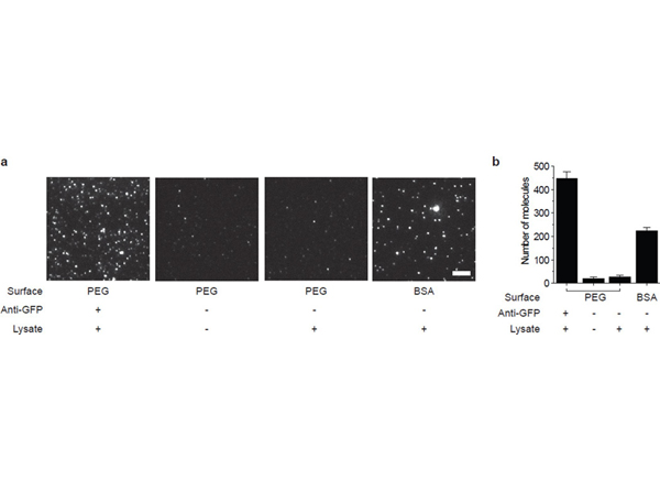
PEG passivation hinders non-specific protein adsorption. (a) Typical TIRF image of YFP-tagged protein pulled down using polyclonal anti-GFP antibody from the lysate (a, left). Non-specific binding of the protein to the PEG passivated surface (a, second from right) is comparable to the blank (a, second from left). Significant non-specific adsorption of YFP is observed when the same amount of lysate is added to surface passivated with 1 mg ml-1?BSA (a, right). Scale bar is 5 μm. (b) Bar graph with average number of fluorophores per image. Error bars represent standard deviation of the mean across > 20 images. Fig 2. PMID:?22322217
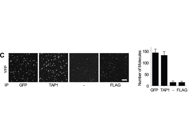
TAP/tapasin ratio is 1:2 in human cells. C, representative fluorescent images (left panel) and quantitation (right panel) of tapasin-YFP pulldown in slides coated with goat anti-GFP, the anti-TAP1 antibody 148.3, no antibody, or a control antibody (mouse anti-FLAG). To achieve an optimal density of molecules for imaging, the .220.B4402.tapasin-YFP lysate was diluted 1000-fold (anti-GFP), 20-fold (anti-TAP1), or 10-fold (no antibody and anti-FLAG) in 1% digitonin lysis buffer. The?scale bar?on the fluorescent images represents 5 μm. The quantitative results are expressed as the means plus the S.D. of at least 20 imaging areas (2500 μm2?each) in one representative experiment.? Fig 1. PMID:?22829594
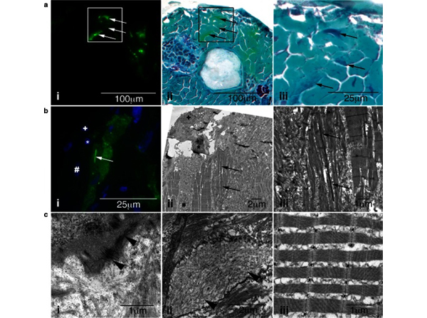
Characterization of skeletal muscle pathology in?Tg(ACTA1?D286G-eGFP)?high?zebrafish.?a?Skeletal muscle expressing?i?mosaic ACTA1D286G-eGFP, and?ii?overlaid with a light microscopy image of the same section showing Gomori trichrome staining, and?iii?enlarged.?Dark regions?(indicative of nemaline bodies) of disrupted muscle correspond to eGFP expression (arrows).?b?Correlative light and electron microscopy of?Tg(ACTA1?D286G-eGFP)?high?fish muscle at 2 dpf.?b?i?Fluorescent image and corresponding?ii?electron microscopy image of skeletal muscle section containing a dense, elongated nemaline body (arrow) and enlarged in (iii). Sections are matched using nuclei positions (asterisk,?plus?and?hash).?c?i?Accumulations of actin filaments (arrowheads) and?ii?diffuse regions of filamentous actin (arrowheads), as well as?ii?disrupted sarcomeric regions are evident in?Tg(ACTA1?D286G-eGFP)?high?skeletal muscle, at 2 dpf unlike the?iii?uniform sarcomeres observed in?Tg(ACTA1-eGFP)?zebrafish. Fig 2. PMID:?25931053
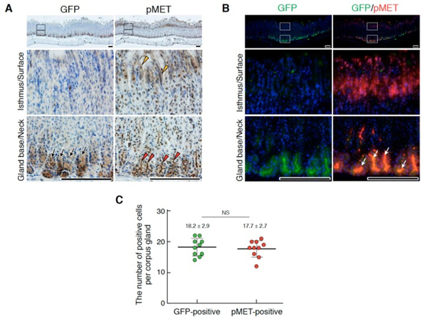
Localizations of Lgr5+?stem cells and pMET in the corpus epithelium of the adult stomach. (A) Immunohistochemical staining for GFP and pMET. (B) Double immunofluorescence staining for GFP and pMET. In (A), black arrows, Lgr5-driven GFP; red arrowheads, pMET in the glandular base region; yellow arrowheads, pMET in the isthmus and/or surface regions. In (B), white arrows indicate the co-localization of pMET and GFP in Lgr5+?stem cells. The images in the lower panel are magnified images of the boxed areas in the upper panel. Tissues were obtained from Lgr5-DTR-EGFP mice. Scale bars represent 200 μm. (C) The numbers of GFP- and pMET-positive cells per corpus gland. These values were obtained by counting in 10 glands (n?= 10). Data were tested for significance using an unpaired two-tailed?t-test. NS, not significant. Fig 6. PMID:?31212972
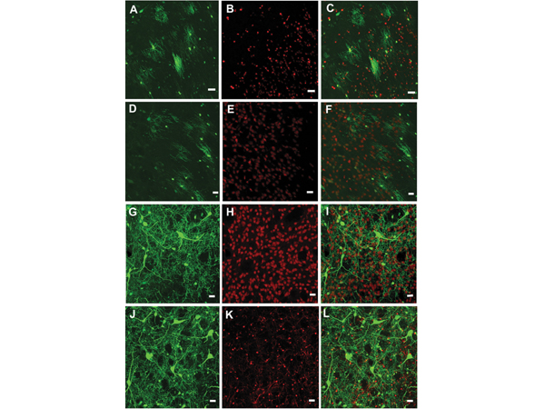
Confocal images of AAV9-hSyn and AAV9AU-hSyn mediated transduction in the rat striatum.?(A?C)?AAV9-JetI-GFP gene expression primarily co-localizes with the oligodendrocyte marker, Olig2, while?(D?F)?the general lack of GFP co-localization is with the neuronal marker, NeuN. In contrast?(G?I),?AAV9AU-JetI-GFP gene expression exhibits substantial co-localization with NeuN, but little co-localization with Olig2. Scale bars equal 20?μm. AAV, adeno-associated virus; GFP, green fluorescent protein. Fig 1. PMID:?32940068
|
|
|
|
ELISA results of purified Goat anti-GST Antibody Biotin Conjugated tested against purified GST. Each well was coated in duplicate with 1.0 μg of GST (p/n 000-001-200). The starting dilution of antibody was 5μg/ml and the X-axis represents the Log10 of a 3-fold dilution. This titration is a 4-parameter curve fit where the IC50 is defined as the titer of the antibody. Assay performed using 3% fish gelatin as blocking buffer, Streptavidin Peroxidase Conjugated and TMB substrate p/n TMBE-1000
|
|
| 別品名 |
goat anti-GST antibody biotin conjugation, biotin conjugated goat anti-GST antibody, Glutathione-S-Transferase, GST antibody, anti-GST antibody, anti-Glutathione-S-Transferase antibody
|
| 適用 |
Enzyme Linked Immunosorbent Assay
|
| 免疫動物 |
Goat
|
| 標識物 |
Biotin
|
| 精製度 |
Affinity Purified
|
| Tag情報 |
GST
|
| 参考文献 |
[[Cosmo Reference Collection]][Pub Med ID]36939020
|
| [注意事項] |
濃度はロットによって異なる可能性があります。メーカーDS及びCoAからご確認ください。
|
|
| メーカー |
品番 |
包装 |
|
RKL
|
600-106-200
|
1 MG
|
※表示価格について
| 当社在庫 |
なし
|
| 納期目安 |
約10日
|
| 法規制 |
毒
|
| 保存温度 |
4℃
|
|
※当社では商品情報の適切な管理に努めておりますが、表示される法規制情報は最新でない可能性があります。
また法規制情報の表示が無いものは、必ずしも法規制に非該当であることを示すものではありません。
商品のお届け前に最新の製品法規制情報をお求めの際はこちらへお問い合わせください。
|
※当社取り扱いの試薬・機器製品および受託サービス・創薬支援サービス(納品物、解析データ等)は、研究用としてのみ販売しております。
人や動物の医療用・臨床診断用・食品用としては、使用しないように、十分ご注意ください。
法規制欄に体外診断用医薬品と記載のものは除きます。
|
|
※リンク先での文献等のダウンロードに際しましては、掲載元の規約遵守をお願いします。
|
|
※CAS Registry Numbers have not been verified by CAS and may be inaccurate.
|