
| 別品名 |
B lymphoma Mo MLV insertion region (mouse) antibody, Bmi 1 antibody, MGC12685 antibody, Murine leukemia viral (bmi 1) oncogene homolog antibody, Oncogene BMI 1 antibody, PCGF 4 antibody
|
| 抗原部位 |
a.a.252-264
|
| 種由来 |
Human
|
| 標識物 |
Unlabeled
|
| 精製度 |
Affinity Purified
|
| 適用 |
Western Blot
Enzyme Linked Immunosorbent Assay
Immuno Fluorescence
|
| 免疫動物 |
Goat
|
| 交差種 |
Human
|
| GENE ID |
648
|
| Accession No.(Gene/Protein) |
P35226
|
| Gene Symbol |
BMI1
|
| 形状 |
滅菌済み液状品
|
| 参考文献 |
Voncken,J.W., Niessen,H., Neufeld,B., Rennefahrt,U., Dahlmans,V., Kubben,N., Holzer,B., Ludwig,S. and Rapp,U.R. (2005) MAPKAP kinase 3pK phosphorylates and regulates chromatin association of the polycomb group protein Bmi1. J. Biol. Chem. 280 (7), 5178-5187. Leung,C., Lingbeek,M., Shakhova,O., Liu,J., Tanger,E., Saremaslani,P., Van Lohuizen,M. and Marino,S. (2004) Bmi1 is essential for cerebellar development and is overexpressed in human medulloblastomas. Nature 428 (6980), 337-341. Obuse,C., Yang,H., Nozaki,N., Goto,S., Okazaki,T. and Yoda,K. (2004) Proteomics analysis of the centromere complex from HeLa interphase cells: UV-damaged DNA binding protein 1 (DDB-1) is a component of the CEN-complex, while BMI-1 is transiently co-localized with the centromeric region in interphase. Genes Cells 9 (2), 105-120.
|
| [注意事項] |
濃度はロットによって異なる可能性があります。メーカーDS及びCoAからご確認ください。
|
|
※サムネイル画像をクリックすると拡大画像が表示されます。
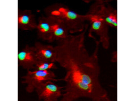
Immunofluorescence using Rockland's affinity purified goat anti Bmi1 shows nuclear staining (green) of methanol fixed (100%, 5 min) HepG2 cells. The cells were blocked and permeabilized in 1%BSA / 10% normal donkey serum / 0.3M glycine in 0.1% PBS Tween for 1h prior to incubation with the primary antibody (1:200 dilution) overnight at +4C and detected with a 488nm fluorescent dye conjugated secondary Ab. Cell nuclei are stained with DAPI (blue) and plasma membranes are stained with WGA (red).
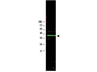
Western blot using Rockland's Affinity Purified anti-Bmi1 antibody shows detection of a band ~37 kDa corresponding to human Bmi1 (arrowhead).?? Approximately 20 ug of a U2OS whole cell lysate (bone osteosarcoma) was separated by 4-20% SDS-PAGE and transferred onto nitrocellulose.? After blocking in PBS containing 5% nonfat dry milk, the membrane was probed overnight at 4 C with the primary antibody diluted to 1:1,000 in PBS containing 1% nonfat dry milk.? The membrane was washed and reacted with a 1:20,000 dilution of IRDyeTM800 conjugated Rb-a-Goat IgG [H&L] MX (605-432-013) for 45 min at room temperature. IRDyeTM800 fluorescence image was captured using the OdysseyR Infrared Imaging System developed by LI-COR. IRDye is a trademark of LI-COR, Inc.? Other detection systems will yield similar results.
|
|
|
|
Immunofluorescence using Rockland's affinity purified goat anti Bmi1 shows nuclear staining (green) of methanol fixed (100%, 5 min) HepG2 cells. The cells were blocked and permeabilized in 1%BSA / 10% normal donkey serum / 0.3M glycine in 0.1% PBS Tween for 1h prior to incubation with the primary antibody (1:200 dilution) overnight at +4C and detected with a 488nm fluorescent dye conjugated secondary Ab. Cell nuclei are stained with DAPI (blue) and plasma membranes are stained with WGA (red).
|
|
|
| メーカー |
品番 |
包装 |
|
RKL
|
600-101-392
|
100 UG
|
※表示価格について
| 当社在庫 |
なし
|
| 納期目安 |
約10日
|
| 保存温度 |
-20℃
|
|

